. 2008 Mar 25;6(3):e67.
doi: 10.1371/journal.pbio.0060067.

Phosphorylation-independent regulation of the diguanylate cyclase WspR

Affiliations

Phosphorylation-independent regulation of the diguanylate cyclase WspR

Nabanita De et al. PLoS Biol. .

Abstract

Environmental signals that trigger bacterial pathogenesis and biofilm formation are mediated by changes in the level of cyclic dimeric guanosine monophosphate (c-di-GMP), a unique eubacterial second messenger. Tight regulation of cellular c-di-GMP concentration is governed by diguanylate cyclases and phosphodiesterases, which are responsible for its production and degradation, respectively. Here, we present the crystal structure of the diguanylate cyclase WspR, a conserved GGDEF domain-containing response regulator in Gram-negative bacteria, bound to c-di-GMP at an inhibitory site. Biochemical analyses revealed that feedback regulation involves the formation of at least three distinct oligomeric states. By switching from an active to a product-inhibited dimer via a tetrameric assembly, WspR utilizes a novel mechanism for modulation of its activity through oligomerization. Moreover, our data suggest that these enzymes can be activated by phosphodiesterases. Thus, in addition to the canonical pathways via phosphorylation of the regulatory domains, both product and enzyme concentration contribute to the coordination of c-di-GMP signaling. A structural comparison reveals resemblance of the oligomeric states to assemblies of GAF domains, widely used regulatory domains in signaling molecules conserved from archaea to mammals, suggesting a similar mechanism of regulation.


Conflict of interest statement

Competing interests. The authors have declared that no competing interests exist.

Figures

Figure 1
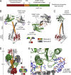
Figure 1. Structure of WspR from P. aeruginosa
(A) Domain organization of WspR. The N-terminal CheY-homology phospho-receiver domain is connected via a helical stalk to the GGDEF domain with diguanylate cyclase activity. In WspR, the active site loop contains the GGEEF motif (residues 251–255). (B) The crystal structure of WspR. The crystals contain two molecules in the asymmetric unit. Two orthogonal views are shown with coloring for molecule A as shown in (A). Molecule B is colored grey. The GGEEF motif is shown in yellow. Cyclic di-GMP molecules bound to the inhibitory site (I-site) are located distal to the active site and are shown as sticks. Mg2+ ions are shown as brown spheres. (C) Comparison of the two WspR molecules in the asymmetric unit. Molecules A and B were aligned through superpositioning of their GGDEF domains. The CheY-stalk modules are separated by a rigid body rotation of 16° around residue 172 at the tip of the helical stalk. (D) Crystallographic tetramer consisting of two symmetry-related dimers representing a biological unit. Two C2-symmetry–related crystallographic dimers of WspR are shown intertwined in a head-to-head orientation. The stalks form a tetrameric structure splaying apart the coiled-coils and physically blocking the active sites. Cyclic di-GMP molecules bound at the I-site bridge the GGDEF domains of neighboring molecules. “S” or “SYM” in the cartoon diagram indicate crystal symmetry-related molecules. The boxed region is shown in (E). (E) Close-up view of the I-site. In the crystal, two intercalated c-di-GMP molecules are bound at the I-site located at the back of the GGDEF domain distal to the catalytic site. An arginine side chain (R198) contributed by a symmetry-related GGDEF domain completes the I-site. Asterisks indicate residues targeted for site-directed mutagenesis.
Figure 2
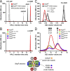
Figure 2. Cyclic di-GMP Binding and Gel Filtration Profile of Wild-Type and Mutant WspR
(A) Detection of guanosine nucleotides by a reverse-phase HPLC-based assay. GTP, GDP, linear di-GMP (pGpG), c-di-GMP, and an intermediate condensation product (linear GTP-GMP; pppGpG) are well separated in this assay (grey dashed line). Products corresponding to pppGpG and pGpG were identified by mass spectrometry (unpublished data). Cyclic di-GMP was commercially available. WspR expressed in E. coli purifies with c-di-GMP bound (red trace) that is accessible for PDEs (black trace). (B) I-site and active site mutants of WspR purify nucleotide-free. Mutant proteins with disrupted I-sites (WspRR242A or WspRR198A; blue and purple traces, respectively) or catalytic site (WspRGGAAF; orange trace) were analyzed. Nucleotide-free WspRwt is obtained by PDE treatment followed by repurification using affinity and size exclusion columns (green trace). (C) PDE treatment triggers a conformational change in WspR. Cyclic di-GMP–bound WspRwt (0.24 mM) was incubated with PDE (0.008 mM) in gel filtration buffer (25 mM Tris-Cl [pH 7.5], 100 mM NaCl, and 1 mM DTT) supplemented with 10 mM Mn2+ for 0.5, 1, or 2 h at 25 °C. Reactions were analyzed by SEC on a Superdex200 10/300 column (GE Heathcare). (D) SEC profiles of mutant and wild-type WspR. Nucleotide-bound and nucleotide-free WspRwt (red and green traces, respectively), WspRGGAAF (orange trace), WspRR242A (blue trace), and WspRR198A (purple trace) (0.24 mM) were analyzed by analytical gel filtration in gel filtration buffer. Peak maxima at 11.7 (peak 1), 12.1 (peak 2), and 13.3 ml (peak 3) are labeled. Peaks 1–3 (Superdex 200 10/30 column; GE Healthcare) correspond to peaks 1–3 (Shodex KW-803 column; JM Science, Inc.) in Table 1, obtained from the SEC coupled to the static multiangle light-scattering detectors.
Figure 5
Figure 5. Reconstitution of a Product-Inhibited WspR
(A) Flow chart outlining the reconstitution experiments. Cyclic di-GMP–bound, inhibited WspRwt (sample 1) (0.24 mM) was treated with PDE (sample 1b). The nucleotide-free, active dimer was repurified (sample 2). After incubation with GTP/Mg2+, the sample was split into two fractions. One fraction was concentrated (to approximately 0.48 mM) and incubated overnight at 4 °C, the other was incubated under identical conditions at its initial concentration (approximately 0.05 mM) (samples 3 and 4, respectively). Samples were subjected to SEC. At each step of the reaction cycle, samples were analyzed for nucleotide content, their gel filtration profile, and enzymatic activity. (B) Nucleotide loading states of WspR species. Proteins were analyzed as described above in Figure 2. (C) Gel filtration profiles of WspR species. WspRwt species indicated in Figure 5A were analyzed as described above (Figure 2C and 2D). Except for the sample that was subjected to a final concentration (sample 4), the concentration of WspR (initially at 0.24 mM) decreased along the reaction scheme due to preparative gel filtration steps. The inset shows concentration-dependent oligomerization behavior of nucleotide-free WspR. WspRwt: nuc.-free (dashed line: 0.08 mM; solid line: 0.24 mM; initial concentration) was analyzed by SEC. Peak maxima are at 11.7 and 13.3 ml elution volume. (D) Enzymatic activity of WspR species. Catalytic activities of WspR species (0.5 μM) were determined in a continuous assay measuring pyrophosphate production as described in Figure 4B. Initial velocities were determined by linear regression. Error bars indicate standard deviations of three independent experiments. (E) Structural and functional characteristics of distinct WspR states. The table summarizes properties of distinct WspR species along the reconstitution path.
Figure 4
Figure 4. Activity of Distinct WspR Species
(A) HPLC-based activity assay measuring c-di-GMP production by WspRwt. Nucleotide-free WspRwt (green trace) or WspRGGAAF (orange trace) (10 μM) was incubated in buffer containing GTP/Mg2+ (1 mM/2 mM) for 1 h at 25 °C. Nucleotides were analyzed by reverse-phase HPLC after heat denaturation of proteins and ultrafiltration of the supernatants, and reaction products were compared to retention times of well-characterized nucleotide standards (grey trace and Figure 2A). (B) Comparison of enzymatic activity of wild-type and mutant forms of WspR. WspRwt (nucleotide-bound or -free), or mutant variants (0.5 μM) were incubated at 25 °C in assay buffer (EnzChek Pyrophosphate Assay; Invitrogen) containing GTP/Mg2+ (0.5 mM/2 mM), and pyrophosphate production was measured by continuously monitoring absorbance at 360 nm. Coloring corresponds to the scheme in Figure 2. Error bars indicate standard deviations of three independent experiments. Incubation of inorganic pyrophosphate (PPi; grey trace) (0.5 mM) in assay buffer determines the rate limit of the assay system. In the buffer control (black trace), GTP/Mg2+ is included in the reaction.
Figure 3
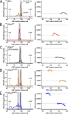
Figure 3. SEC-Coupled Multiangle Light-Scattering Analysis of Purified WspR in Solution
(A) Monomeric BSA (4 mg/ml; Sigma) was analyzed by coupled SEC/multiangle light scattering. The mobile phase consists of 25 mM Tris-HCl (pH 7.5), 100 mM NaCl, and 1 mM DTT. In the left panel, the primary signal (in volts) is plotted against the elution volume. The solid (colored) trace shows the signal of one of the light-scattering detectors (at 90° to the incident beam). The signal from the refractive index detector is shown as a dashed line. The grey area highlights the analyzed peak. The void volume (void) and the end of the experiment (buffer) are indicated. In the right panel, molecular weights, determined by light scattering, and protein concentration, measured by change of refractive index, from each data slice (0.5-s increments) are plotted against the elution volume. The grey horizontal line indicates the theoretical molecular weight. (B–E) Cyclic di-GMP–bound WspRwt (4 mg/ml) (B), PDE-treated WspRwt (4 mg/ml) (C), WspRGGAAF (4 mg/ml) (D), and WspRR242A (4 mg/ml) (E) were analyzed as described in (A). The grey horizontal lines indicate the theoretical molecular weight for monomers, dimers, and tetramers.
Figure 6
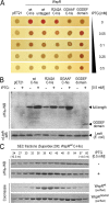
Figure 6. Catalytic Activity and Oligomerization of WspR in Cells
(A) Congo Red (CR) assay monitoring WspR-catalyzed c-di-GMP production in cells. E. coli BL21 were transformed with plasmids encoding wild-type or mutant variants of WspR. Cells were grown to mid-log phase at 37 °C, and 2.5 μl of the culture was spotted onto a CR-containing LB plate with or without IPTG and incubated for 24 h at 30 °C. Leaky expression in the absence of IPTG and IPTG-induced WspR expression was visually assayed for a red colony phenotype (rdar morphotype). Cells expressing an untagged version of WspRwt behave similarly to cells expressing WspR with a C-terminal hexahistidine tag. (B) Loss of CR staining correlates with high WspR expression levels. Cultures were grown for 16 h at 25 °C in the absence or presence of IPTG. Lysates from cells expressing hexahistidine-tagged wild-type and mutant variants of WspR (see above) were prepared by sonication and analyzed by western blotting using a hexahistidine tag-specific antibody to detect recombinant protein. Samples were normalized to total protein amount prior to SDS-PAGE and blotting. Western blot detection of the native E. coli protein LexA was used as a control. LexA levels were slightly lower in some samples due to high WspR expression levels obscuring total protein normalization. (C) Gel filtration profile of WspR in cell lysates. Cell lysates were subjected to SEC in gel filtration buffer. Fractions (0.1 ml) were collected, and hexahistidine-tagged proteins in the fractions were detected by western blotting. Elution profiles were compared to profiles obtained for purified proteins (c-di-GMP–bound WsrRwt or nucleotide-free WspRGGAAF) under identical conditions.
Figure 7
Figure 7. Model for the Feedback Regulation and Reactivation of WspR
Based on structural and functional analyses, a model is proposed in which active WspR dimers are in equilibrium with a transient tetrameric species that, in the presence of c-di-GMP, provides an exit platform for the product-inhibited elongated dimer. Degradation of c-di-GMP by PDEs triggers a snapping back to the active dimer species, probably avoiding the tetrameric state. Both c-di-GMP binding and tetramerization are required for the assembly of the inhibited species. It is unlikely that the tetrameric state is en route from the inhibited to the active dimer, which would require dissociation of the tetramer once c-di-GMP has been degraded. Experimentally, we did not observe any tetramers immediately after PDE treatment, and the tetrameric species arises only after prolonged incubation of the compact, but not the elongated dimer (Table 1, see also Figures 2C, 3, and 5).
Figure 8
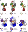
Figure 8. Distinct Oligomeric Conformations of WspR and Mechanistic Implication for the Regulation of GAF Domain-Containing Proteins
(A) Structural models closely resembling the distinct states of WspR. The solvent-accessible surface of a WspR dimer is shown. In the active state (left panel), dimerization is mediated by the CheY-homology domains and protruding stalks. The stalks may convene in the fully active state bringing the GGDEF domains into close proximity (arrow in left panel). In the tetrameric assembly that serves as an intermediate between the two dimeric states (middle panel), two C2-symmetry–related crystallographic dimers of WspR are shown intertwined in a head-to-head orientation. “S” or “SYM” in the cartoon diagram indicates crystal symmetry–related molecules. The stalks form a tetrameric structure splaying apart the coiled-coils and physically blocking the active sites. Cyclic di-GMP molecules bound at the I-site bridge the GGDEF domains of neighboring molecules. Arrows indicate how breaking up the CheY domain dimer by a rigid body rotation of the CheY-stalk module would facilitate the formation of two identical dimers shown in the right panel. In the proposed model for the product-inhibited state, dimers of two symmetry-related molecules (chain A) are held together by three interfaces between the tip of the stalks (zone 1), between the GGDEF domain and the stalk (zone 2), and between the GGDEF domain and the CheY domain (zone 3) of adjacent molecules. Such dimers can be readily obtained from the tetramer by segregation of a plane consisting of the colored molecules (chain A dimer) from a plane formed by the grey molecules (chain B dimer). The maximal dimensions and surface areas buried at the interfaces are shown. (B) Dimeric and tetrameric assembly seen in the mouse PDE2A tandem GAF domain crystal structure. The molecule in the asymmetric unit and a symmetry-related molecule (indicated by apostrophes) form a dimer via pairing of their GAF A domains (PDB code 1MC0) [39]. In the crystal, the tips of the stalks that connect the GAF A and GAF B domains are splayed apart by a symmetry-related dimer (see tetrameric structure). Cyclic GMP is bound only to the GAF B domains. The tetrameric assembly consists of two additional symmetry mates (indicated by asterisks). The two symmetry-related crystallographic dimers are intertwined in a head-to-head orientation with the stalks forming a tetrameric structure via their split ends. In such an assembly, significant interfaces for the dimer–dimer interaction are formed between the stalks, between the GAF B domains, and between the GAF A and B domains of adjacent chains. GAF A domains are colored blue and light blue, GAF B domains are colored violet and light violet, and the stalks are shown in orange and grey. (C) Crystal structure of the tandem GAF domain dimer from the adenylate cyclase cyaB2 in Anabaena. The two tandem GAF domain molecules in the asymmetric unit form an antiparallel dimer mediated in part by the helical stalks connecting the GAF A and B domains (PDB code 1YKD) [38]. Cyclic AMP is bound to both GAF A and B domains of cyaB2. The coloring scheme in (B) has been applied for straightforward comparison of relative domain orientations.

Comment in

Similar articles

Cited by

References

    1. Stock AM, Robinson VL, Goudreau PN. Two-component signal transduction. Annu Rev Biochem. 2000;69:183–215. - PubMed
    1. Galperin M. Structural classification of bacterial response regulators: diversity of output domains and domain combinations. J Bacteriol. 2006;188:4169–4182. - PMC - PubMed
    1. D'Argenio D, Miller S. Cyclic di-GMP as a bacterial second messenger. Microbiology. 2004;150:2497–2502. - PubMed
    1. Römling U, Gomelsky M, Galperin M. C-di-GMP: the dawning of a novel bacterial signalling system. Mol Microbiol. 2005;57:629–639. - PubMed
    1. Ross P, Mayer R, Weinhouse H, Amikam D, Huggirat Y, et al. The cyclic diguanylic acid regulatory system of cellulose synthesis in Acetobacter xylinum. Chemical synthesis and biological activity of cyclic nucleotide dimer, trimer, and phosphothioate derivatives. J Biol Chem. 1990;265:18933–18943. - PubMed

Publication types

MeSH terms

Associated data

LinkOut - more resources