. 2008 Jan-Feb;12(1):271-80.
doi: 10.1111/j.1582-4934.2007.00119.x.

Isolation of human foetal myoblasts and its application for microencapsulation

Affiliations

Isolation of human foetal myoblasts and its application for microencapsulation

Anna Aihua Li et al. J Cell Mol Med. 2008 Jan-Feb.

Abstract

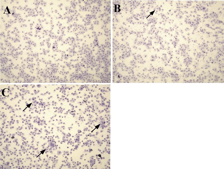
Foetal cells secrete more growth factors, generate less immune response, grow and proliferate better than adult cells. These characteristics make them desirable for recombinant modification and use in microencapsulated cellular gene therapeutics. We have established a system in vitro to obtain a pure population of primary human foetal myoblasts under several rounds of selection with non-collagen coated plates and identified by desmin staining. These primary myoblasts presented good proliferation ability and better differentiation characteristics in monolayer and after microencapsulation compared to murine myoblast C2C12 cells based on creatine phosphokinase (CPK), major histocompatibility complex (MHC) and multi-nucleated myotubule determination. The lifespan of primary myoblasts was 70 population doublings before entering into senescent state, with a population time of 18-24 hrs. Hence, we have developed a protocol for isolating human foetal primary myoblasts with excellent differentiation potential and robust growth and longevity. They should be useful for cell-based therapy in human clinical applications with microencapsulation technology.


Figures

1
1
Myogenic enrichment examined by desmin staining. (A) Fibroblasts at passage 2 under phase contrast microscopy; (B) The same fibroblasts at passage 2 under light transmission microscopy; (C) C2C12 cell line; (D) Human primary myoblasts at passage 5. All the cells were grown for 24 hrs, fixed with 95% methanol and stained with anti-desmin antibody. (Magnification: × 160)
2
2
Creatine phosphokinase (CPK) activity in differentiation medium. The cells were cultured in growth medium for 24 hrs and switched into differentiation medium. The cells were trypsinized and pelleted by centrifugation at different time points. The pellets were suspended in phosphate buffered saline (PBS) and sonicated to release the cytoplasm. The CPK was assayed and standardized by protein concentration.
3
3
Expression of major histocom-patibility complex (MHC). The cells were cultured in growth medium for 24 hrs and switched into differentiation medium. At different time points, the cells were fixed with 95% methanol and stained with anti-MHC antibody. (A) Human foetal primary fibroblasts with no positive stain; (B) C2C12 cells were differentiated into myotubes, but with lots of dead cells; (C) Human foetal primary myoblasts demonstrated high expression of MHC and multi-nucleated myotubes at day 2 with few dead cells; (D) human foetal primary myoblasts remained in well-differentiation state at day 12 with less dead cells. (Magnification: × 160)
4
4
Multi-nucleated cells stained by Giemsa.(A) Human foetal primary fibroblasts; (B) C2C12 cells; (C) Human foetal primary myoblasts. The cells were cultured in growth medium for 24 hrs and transferred into differentiation medium. At day 9, the cells were trypsinized and centrifuged onto slides. The cells on the slides were fixed in 95% methanol and stained in Giemsa solution.(Magnification: × 160)
5
5
Comparison of growth and differentiation of encapsulated foetal human myoblasts and C2C12 cells. Encapsulated cells were grown in growth media for 2 weeks and transferred to differentiation media and sampled at different time points for growth (A), CPK activity (B), MHC expression (C) and myogenic index (D).

References

    1. Emmrich F, Keitel R, Sorger K, Otto U, Staffa G, Klotzer B. Quantitative alterations of immune serum globulin concentrations in pigs transplanted with a renal allograft. Exp Pathol. 1977;14:334–9. - PubMed
    1. Husberg BS. The influence of cyclophosphamide on the rejection of allogenic rat kidney transplants. An in vitro study of cell-bound and antibody-mediated immunity. Clin Exp Immunol. 1972;10:697–711. - PMC - PubMed
    1. Foglia RP, DiPreta J, Statter MB, Donahoe PK. Fetal allograft survival in immunocompetent recipients is age dependent and organ specific. Ann Surg. 1986;204:402–10. - PMC - PubMed
    1. Statter MB, Fahrner KJ, Barksdale EM, Jr, Parks DE, Flavell RA, Donahoe PK. Correlation of fetal kidney and testis congenic graft survival with reduced major histocompatibility complex burden. Transplantation. 1989;47:651–60. - PubMed
    1. Nouwen EJ, Dauwe S, Van Der Biest I, De Broe ME. Stage- and segment-specific expression of cell-adhesion molecules N-CAM, A-CAM, and L-CAM in the kidney. Kidney Int. 1993;44:147–58. - PubMed