. 2008 Jan-Feb;12(1):281-91.
doi: 10.1111/j.1582-4934.2007.00103.x.

Calcium phosphate surfaces promote osteogenic differentiation of mesenchymal stem cells

Affiliations

Calcium phosphate surfaces promote osteogenic differentiation of mesenchymal stem cells

Petra Müller et al. J Cell Mol Med. 2008 Jan-Feb.

Abstract

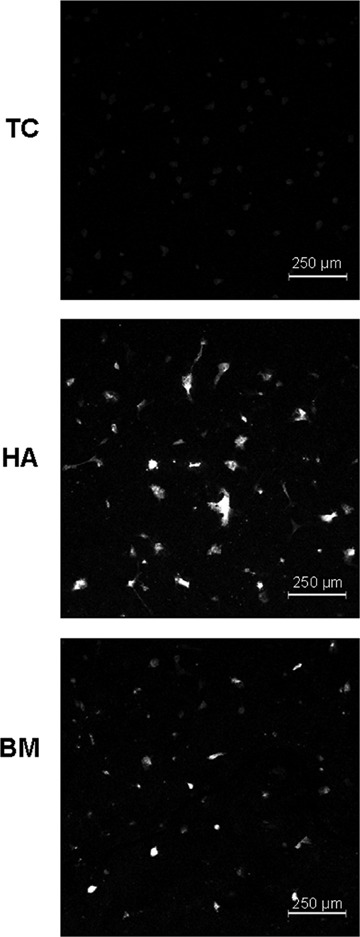
Although studies in vivo revealed promising results in bone regeneration after implantation of scaffolds together with osteogenic progenitor cells, basic questions remain how material surfaces control the biology of mesenchymal stem cells (MSC). We used human MSC derived from bone marrow and studied the osteogenic differentiation on calcium phosphate surfaces. In osteogenic differentiation medium MSC differentiated to osteoblasts on hydroxyapatite and BONITmatrix, a degradable xerogel composite, within 14 days. Cells revealed a higher alkaline phosphatase (ALP) activity and increased RNA expression of collagen I and osteocalcin using real-time RTPCR compared with cells on tissue culture plastic. To test whether material surface characteristics alone are able to stimulate osteogenic differentiation, MSC were cultured on the materials in expansion medium without soluble additives for osteogenic differentiation. Indeed, cells on calcium phosphate without osteogenic differentiation additives developed to osteoblasts as shown by increased ALP activity and expression of osteogenic genes, which was not the case on tissue culture plastic. Because we reasoned that the stimulating effect on osteogenesis by calcium phosphate surfaces depends on an altered cell-extracellular matrix interaction we studied the dynamic behaviour of focal adhesions using cells transfected with GFP labelled vinculin. On BONITmatrix, an increased mobility of focal adhesions was observed compared with cells on tissue culture plastic. In conclusion, calcium phosphate surfaces are able to drive MSC to osteoblasts in the absence of osteogenic differentiation supplements in the medium. An altered dynamic behaviour of focal adhesions on calcium phosphate surfaces might be involved in the molecular mechanisms which promote osteogenic differentiation.


Figures

1
1
Scanning electron microscopy of mesenchymal stem cells on tissue culture plastic (a), hydroxyapatite (b) and BONITmatrix® (c). On all three surfaces, cells reveal a similar flat morphology.
2
2
ALP activity measured in cells on tissue culture plastic (TC), hydroxyapatide (HA) and BONITmatrix® (BM) after mesenchymal stem cells were cultured for 14 days in ODM on the different substrates. Results reveal an increased ALP activity on calcium phosphate surfaces.
3
3
RNA expression of ALP, collagen I (Col1), bone sialoprotein (BSP), osteocalcin (OCN) and transcription factor Runx2 in cells, after mesenchymal stem cells (MSC) were cultured 3 days and 14 days in ODM on tissue culture plastic (black columns) and on BONITmatrix® (grey columns). Results were related to controls (dashed line) representing MSC cultured on tissue culture plastic in EM. For collagen I (on day 3), osteocalcin (on day 14) and Runx2 (both days) an increased expression on BONITmatrix® was observed. (Asterix indicates statistically significant difference between cells on the two different materials.)
4
4
(a) Fluorescence images of ALP activity in cells on tissue culture plastic (TC), hydroxyapatite (HA) and BONITmatrix® after mesenchymal stem cells were cultured in EM on different substrates for 14 days. Note that there was no visible ALP activity in cells on TC. (b) Quantitative analyses of ALP activity in cells on TC, HA and BM. The highest ALP activity was measured in cells on HA.
5
5
Quantitative analyses of ALP activity in MG-63 cells on TC, HA and BM after cells were cultured for 7 days in EM (black columns) or ODM (grey columns). ALP activity was only found in cells on calcium phosphate surfaces both in ODM and EM.
6
6
RNA expression of ALP, Col1, BSP, OCN, and Runx2 after mesenchymal stem cells were cultured for 3 and 14 days in EM on BONITmatrix®. Results were related to controls (dashed line) representing cells cultured in EM on tissue culture plastic.
7
7
RNA expression of ALP after MG-63 cells were cultured for 3 days and 7 days on tissue culture plastic (TC/3 and TC/7, respectively) and 3 and 7 days on BONITmatrix® (MB/3 and BM/7, respectively). Cells were cultured both in EM (black columns) and in ODM (grey columns). Results were related to controls (dashed line) representing cells cultured in EM on TC. On BM, MG-63 cells revealed an increased expression of ALP.
8
8
Analyses of focal adhesions in live MG-63 cells on different materials after transfection of GFP-vinculin. (A) Number of focal adhesions per cell: Lower number of focal adhesions in cells on BONITmatrix® (BM) compared with cells on collagen coated chamber slides (Col) and on titanium (Ti). (B) Size of focal adhesions:Lower size of focal adhesions in cells on BM compared with cells on Col and Ti. (C) Speed of the movement of focal adhesions:Increased mobility of focal adhesions in cells on BM compared with Col and Ti, where the lowest movement was observed.(Asterix indicates statistically significant differences between cells on BM compared with cells on the two other materials.)

Similar articles

Cited by

References

    1. Salgado AJ, Coutinho OP, Reis RL. Bone tissue engineering: state of the art and future trends. Macromol Biosc. 2004;4:743–65. - PubMed
    1. Otto WR, Rao J. Tomorrow's skeleton staff: mesenchymal stem cells and the repair of bone and cartilage. Cell Prolif. 2004;37:97–110. - PMC - PubMed
    1. Pountos I, Jones E, Tzioupis C, McGonagle D, Giannoudis PV. Growing bone and cartilage. The role of mesenchymal stem cells. J Bone Joint Surg Br. 2006;88:421–6. - PubMed
    1. Petite H, Viateau V, Bensaid W, Meunier A, De Pollak C, Bourguignon M, Oudina K, Sedel L, Guillemin G. Tissue-engineered bone regeneration. Nat Biotechnol. 2000;18:959–63. - PubMed
    1. Mistry AS, Mikos AG. Tissue engineering strategies for bone regeneration. Adv Biochem Eng Biotechnol. 2005;94:1–22. - PubMed

Publication types