. 2008;10(2):R25.
doi: 10.1186/bcr1982. Epub 2008 Mar 26.

Human breast cancer cell lines contain stem-like cells that self-renew, give rise to phenotypically diverse progeny and survive chemotherapy

Affiliations

Human breast cancer cell lines contain stem-like cells that self-renew, give rise to phenotypically diverse progeny and survive chemotherapy

Christine M Fillmore et al. Breast Cancer Res. 2008.

Abstract

Introduction: The phenotypic and functional differences between cells that initiate human breast tumors (cancer stem cells) and those that comprise the tumor bulk are difficult to study using only primary tumor tissue. We embarked on this study hypothesizing that breast cancer cell lines would contain analogous hierarchical differentiation programs to those found in primary breast tumors.

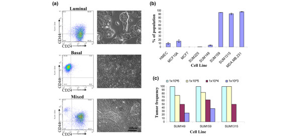
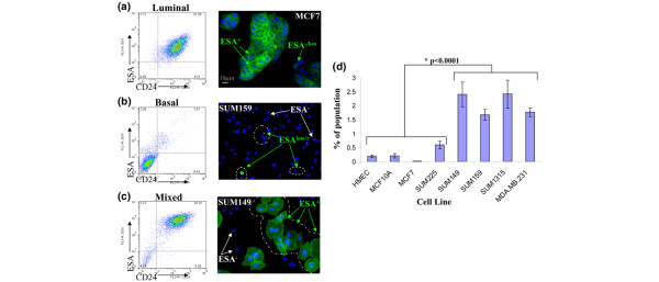
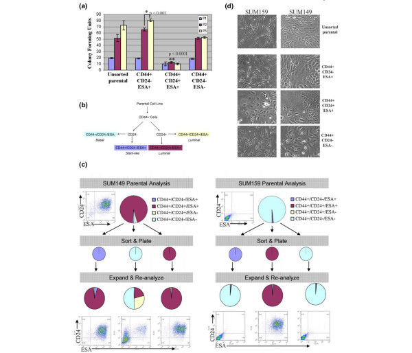
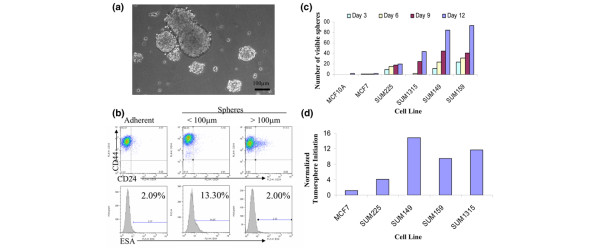
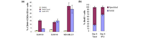
Methods: Eight human breast cell lines (human mammary epithelial cells, and MCF10A, MCF7, SUM149, SUM159, SUM1315 and MDA.MB.231 cells) were analyzed using flow cytometry for CD44, CD24, and epithelial-specific antigen (ESA) expression. Limiting dilution orthotopic injections were used to evaluate tumor initiation, while serial colony-forming unit, reconstitution and tumorsphere assays were performed to assess self-renewal and differentiation. Pulse-chase bromodeoxyuridine (5-bromo-2-deoxyuridine [BrdU]) labeling was used to examine cell cycle and label-retention of cancer stem cells. Cells were treated with paclitaxel and 5-fluorouracil to test selective resistance to chemotherapy, and gene expression profile after chemotherapy were examined.

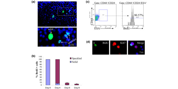
Results: The percentage of CD44+/CD24- cells within cell lines does not correlate with tumorigenicity, but as few as 100 cells can form tumors when sorted for CD44+/CD24-/low/ESA+. Furthermore, CD44+/CD24-/ESA+ cells can self-renew, reconstitute the parental cell line, retain BrdU label, and preferentially survive chemotherapy.

Conclusion: These data validate the use of cancer cell lines as models for the development and testing of novel therapeutics aimed at eradicating cancer stem cells.


Figures

Figure 1
Figure 1
CD44+/CD24- cells associate with a basal phenotype not with tumorigenic potential of human breast cancer cell lines. (a) Luminal, basal or mixed cell morphology can be predicted based on CD44 and CD24 expression. Provided are representative flow cytometry dot plots of CD44 and CD24 expression adjacent to phase-contrast brightfield images of each cell line subtype. Cell lines are composed of luminal-like, CD24+ expressing cells (MCF7 and SUM225); basal-like, CD24- expressing cells (SUM159, SUM1315, and MDA.MB.231); or mixed luminal and basal-like cells and consist of both CD24+ and CD24- populations (human mammary epithelial cells [HMEC] and SUM149). (b) The average percentage of CD44+/CD24- cells in each cell line was determined by flow cytometry. Data are presented as the mean ± standard error of the mean for four separate experiments. (c) The frequency of tumor growth was measured for SUM149, SUM159, and SUM1315 cell lines injected orthotopically in nonobese diabetic/severe combined immunodeficient mice when a tumor was palpable (>3 mm) within 100 to 150 days.
Figure 2
Figure 2
ESA can distinguish luminal, basal, and stem-like cells. Presented are representative flow cytometry dot plots of epithelial-specific antigen (ESA) and CD24 expression adjacent to ESA immunofluorescence of each cell line subtype. Cell lines are as follows: (a) luminal-like, expressing high levels of ESA and CD24 (MCF7, SUM225, and T47D); (b) basal-like, expressing low levels of ESA and CD24 (SUM159, SUM1315, and MDA.MB.231); or (c) mixed luminal/basal cultures that contain both ESA+/CD24+ and ESA-/CD24- populations (SUM149). (d) The average percentage of CD44+/CD24-/ESA+ cells in each cell line, as determined by flow cytometry. Data are presented as the mean ± stamdard error of the mean for five separate experiments. HMEC, human mammary epithelial cells.
Figure 3
Figure 3
CD44+/CD24-/low/ESA+ cells in human breast cancer cell lines enrich for tumorigenicity. Shown are representative flow cytometry analysis and gates for epithelial-specific antigen (ESA), CD24 and CD44 sorting. (a) SUM159 cells were sorted on ESA. Dot plots show resulting CD44+/CD24-/low/ESA+ or CD44+/CD24-/ESA- fractions. (b) SUM149 cells were sorted on CD44 and CD24 and subsequently sorted for ESA+, shown in histograms. (c) Histological analysis of SUM159 and SUM149 orthotopic tumors. The upper panels present representative hematoxylin and eosin stained sections at 20× magnification of tumors that formed from 5 × 10⁵ unsorted, 1,000 CD44+/CD24-/low/ESA+, or 1 × 10⁶ CD44+/CD24-/ESA- SUM159 cells. Note that the tumor histology from CD44+/CD24-/ESA- sorted cells is different than that from unsorted or CD44+/CD24-/low/ESA+ cells. The lower panels present hematoxylin and eosin stained sections at 20× magnification of tumors that formed from 1 × 10⁵ unsorted, CD44+/CD24-/low/ESA+, or CD44+/CD24+/ESA+ sorted SUM149 cells. Note the tumor histology from CD44+/CD24+/ESA+ sorted cells is markedly different from that of the unsorted or the CD44+/CD24-/low/ESA+ sorted cells.
Figure 4
Figure 4
CD44+/CD24-/ESA+ cells are enriched for self-renewal activity in vitro. (a) Serial colony-forming units (CFUs) from 300 SUM159 sorted cells. Data are present as mean ± standard error of the mean for three separate experiments. Exhaustion of CFU is defined as the P3 colonies = P2 colonies. (b) Flow chart describing sorting scheme for reconstitution. (c) SUM149 or SUM159 sorted cells were expanded and the cellular outgrowths were reanalyzed by flow cytometry to assay reconstitution of the parental line. The parental line was run through the flow cytometer and expanded in parallel to control of the effects of fluorescence-activated cell sorting (top).(d) Representative phase-contrast brightfield images of unsorted SUM159 or SUM149, and colonies that grew out from CD44+/CD24+/ESA+, CD44+/CD24+/ESA-, CD44+/CD24-/ESA+, or CD44+/CD24-/ESA- sorted cells. ESA, epithelial-specific antigen.
Figure 5
Figure 5
Tumorsphere formation correlates with presence of CD44+/CD24-/ESA+ cells. (a) Phase-contrast brightfield image of SUM159 tumorspheres at day 11 under a 10× objective. (b) Representative flow cytometry profiles of SUM159 cells grown in adherent or nonadherent cultures. Nonadherent tumorspheres were filtered through cell strainers to separate spheres smaller than 100 μm from spheres larger than 100 μm. Percentage indicated in epithelial-specific antigen (ESA) histograms represents the population of CD44+/CD24-/ESA+ cells. (c) For each cell line, 5,600 single cells/cm2 were plated on super-low adherence plates and spheres visible by eye were counted every 3 days. (d) The absolute number of tumorspheres on day 12 was divided by the corresponding crystal violet value (see Materials and methods) for each cell line to correct for the proliferation rate of the cell lines.
Figure 6
Figure 6
CD44+/CD24-/ESA+ cells retain BrdU label. (a) SUM159 were continuously treated with 5-bromo-2-deoxyuridine (BrdU) for 10 days. Cells that stain for BrdU label 6 days after chase exhibit either a solid or speckled pattern of label retention: BrdU (green) or DAPI (4',6 diamidino-2-phenylindole; blue). (b) SUM159 and MDA.MB.231 cells were pulse-chased with BrdU. The percentage of cells with either solid or speckled BrdU staining were counted at the indicated time points. Data are presented as the mean ± standard error of the mean for at least three separate 10× fields of two separate experiments. (c) Proliferating cultures of SUM159 cells were pulse-chased and analyzed by flow cytometry for CD44+/CD24-/ESA+ and the presence of BrdU. Note that about 90% of CD44+/CD24-/ESA+ SUM159 cells retain BrdU label at 6 days after chase. (d) Immunoflourescence of BrdU and Ki67 in cells that were BrdU pulse-chased. (i) Co-localization of BrdU (green) and Ki67 (red); note that of the adjacent nuclei that are both Ki67 positive, only one contains BrdU. ESA, epithelial-specific antigen.
Figure 7
Figure 7
CD44+/CD24-/ESA+ cells preferentially survive chemotherapy. (a) Quantification of viable CD44+/CD24-/ESA+ cells in various human breast cancer cell lines following a 6-day treatment with 10 nmol/l paclitaxol (Taxol) or 1 mm 5-fluorouracil (5-FU). Data are presented as the mean ± standard error of the mean for four separate experiments. (b) SUM159 cells were continuously treated with BrdU for 10 days and chased with media containing 10 nmol/l Taxol or 1 mmol/l 5-FU. On day 6 of the chase, cells were stained for 5-bromo-2-deoxyuridine (BrdU) and counted for the solid (complete label retention) and speckled (partial label retention) staining. DMSO, dimethyl sulfoxide.

References

    1. Dvoark H, Flier J, Frank H. Tumours – wounds that do not heal: similarities between tumour stroma generation and wound healing. N Engl J Med. 1986;315:1650–1659. - PubMed
    1. Al-Hajj M, Wicha SM, Benito-Hernandez A, Morrison SJ, Clarke Mf. Prospective identification of tumorigenic breast cancer cells. Proc Natl Acad Sci USA. 2003;100:3983–3988. doi: 10.1073/pnas.0530291100. - DOI - PMC - PubMed
    1. Collins AT, Berry PA, Hyde C, Stower MJ, Maitland NJ. Prospective identification of tumorigenic prostate cancer stem cells. Cancer Res. 2005;65:10946–10951. doi: 10.1158/0008-5472.CAN-05-2018. - DOI - PubMed
    1. O'Brien CA, Pollet A, Gallinger S, Dick JE. A human colon caner cell capable of initiating tumour growth in immunodeficient mice. Nature. 2007;445:106–110. doi: 10.1038/nature05372. - DOI - PubMed
    1. Ricci-Vitiani L, Lonbardi DG, Pilozzi E, Biffoni M, Todaro M, Peschle C, De Maria R. Identification and expansion of human colon cancer-initiating cells. Nature. 2007;445:111–115. doi: 10.1038/nature05384. - DOI - PubMed

Publication types

MeSH terms