. 2008 Mar 13:9:29.
doi: 10.1186/1471-2199-9-29.

Association of ATRX with pericentric heterochromatin and the Y chromosome of neonatal mouse spermatogonia

Affiliations

Association of ATRX with pericentric heterochromatin and the Y chromosome of neonatal mouse spermatogonia

Claudia Baumann et al. BMC Mol Biol. .

Abstract

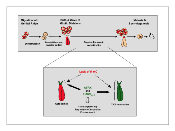
Background: Establishment of chromosomal cytosine methylation and histone methylation patterns are critical epigenetic modifications required for heterochromatin formation in the mammalian genome. However, the nature of the primary signal(s) targeting DNA methylation at specific genomic regions is not clear. Notably, whether histone methylation and/or chromatin remodeling proteins play a role in the establishment of DNA methylation during gametogenesis is not known. The chromosomes of mouse neonatal spermatogonia display a unique pattern of 5-methyl cytosine staining whereby centromeric heterochromatin is hypo-methylated whereas chromatids are strongly methylated. Thus, in order to gain some insight into the relationship between global DNA and histone methylation in the germ line we have used neonatal spermatogonia as a model to determine whether these unique chromosomal DNA methylation patterns are also reflected by concomitant changes in histone methylation.

Results: Our results demonstrate that histone H3 tri-methylated at lysine 9 (H3K9me3), a hallmark of constitutive heterochromatin, as well as the chromatin remodeling protein ATRX remained associated with pericentric heterochromatin regions in spite of their extensive hypo-methylation. This suggests that in neonatal spermatogonia, chromosomal 5-methyl cytosine patterns are regulated independently of changes in histone methylation, potentially reflecting a crucial mechanism to maintain pericentric heterochromatin silencing. Furthermore, chromatin immunoprecipitation and fluorescence in situ hybridization, revealed that ATRX as well as H3K9me3 associate with Y chromosome-specific DNA sequences and decorate both arms of the Y chromosome, suggesting a possible role in heterochromatinization and the predominant transcriptional quiescence of this chromosome during spermatogenesis.

Conclusion: These results are consistent with a role for histone modifications and chromatin remodeling proteins such as ATRX in maintaining transcriptional repression at constitutive heterochromatin domains in the absence of 5-methyl cytosine and provide evidence suggesting that the establishment and/or maintenance of repressive histone and chromatin modifications at pericentric heterochromatin following genome-wide epigenetic reprogramming in the germ line may precede the establishment of chromosomal 5-methyl cytosine patterns as a genomic silencing strategy in neonatal spermatogonia.


Figures

Figure 1
Figure 1
Comparison of Global DNA methylation patterns in the chromosomes of mouse embryonic fibroblasts and neonatal spermatogonia. A-C) Fluorescent micrographs of metaphase chromosomes from primary mouse embryonic fibroblasts showing highly methylated autosomal centromeric domains (thin arrow) as determined by 5-methyl cytosine (5-mC) staining (red). D-F) Chromosomes in neonatal spermatogonia lack global DNA methylation at pericentric heterochromatin, while 5-mC (red) decorates entire chromatids in the majority of autosomes (thin arrow). Two chromosomes on each metaphase spread consistently exhibit lack of 5-mC staining (bold arrow and arrowhead). These patterns are reproducible after using two standard chromosome fixation protocols using either paraformaldehyde (PFA) or methanol/acetic acid (MeOH/AA) as shown in (G-I). J-L) Fluorescent in situ hybridization (FISH) and subsequent determination of DNA methylation patterns (5-mC; red) confirms that the two hypo-methylated chromosomes observed on each spread correspond to the X chromosome (green; arrow head) and the Y chromosome (red; Inset and bold arrow in K-L). Note the lack of 5-mC staining on the Y chromosome and the low levels of global methylation on the X chromosome compared to autosomes (thin arrows) in germ cell metaphases. DNA was counterstained with HOECHST 33258 (blue). Scale bars = 10 μm.
Figure 2
Figure 2
Chromosomal localization of ATRX and histone H3 tri-methylation on lysine 9 (H3K9me3) in neonatal spermatogonia. A) Analysis of histone methylation patterns revealed that in spite of the absence of global DNA methylation (red) at pericentric heterochromatin (thin arrow), H3K9me3 (green) remained associated with centromeric domains in the chromosomes of neonatal spermatogonia. Importantly, H3K9me3 is also preferentially associated with one of the unmethylated chromosomes on each spread analyzed (bold arrow). B) In contrast, H3K4me2 (green) remains associated with entire chromatids in the majority of chromosomes (thin arrow). However, one of the unmethylated chromosomes was consistently found to be completely devoid of H3K4me2 (bold arrow). C) Notably, the chromatin remodeling protein ATRX (green) remains associated with pericentric heterochromatin and consistently marks a single unmethylated chromosome on each metaphase spread analyzed. Scale bar = 10 μm.
Figure 3
Figure 3
Repressive histone and chromatin modifications in the absence of global DNA methylation in the Y chromosome of neonatal spermatogonia. A-C) Immuno-FISH analysis of neonatal spermatogonia revealed that the ATRX protein (green) as well as H3K9me3 (green; D-F) consistently associate with pericentric heterochromatin domains in autosomes (thin arrows). In addition to their centromeric localization, ATRX as well as H3K9me3 preferentially associate with both arms of the Y chromosome (red; bold arrows). G-I) In contrast, a transcriptionally permissive histone modification such as H3K4me2 (green) exhibited an inverted chromosomal distribution whereby constitutive heterochromatin (i.e. pericentric heterochromatin and the Y chromosome; red) are completely devoid of H3K4me2. The Y chromosome in neonatal spermatogonia occupies a transcriptionally quiescent nuclear domain during interphase. Transcription run-on assays after incorporation of Br-UTP (green) in permeabilized interphase nuclei of somatic (J-L) and neonatal spermatogonia (M-O) revealed that global transcriptional activity is undetectable in the nuclear domain occupied by the Y chromosome (red; see inset) in both somatic cell and germ cell nuclei. Neonatal spermatogonial cell nuclei were identified by their unique global DNA methylation patterns (5-mC; green). Scale bar = 10 μm.
Figure 4
Figure 4
Specific association of ATRX and H3K9me3 with lack of H3K4me2 at repetitive DNA sequences on the Y chromosome. A) Metaphase spreads of peripheral lymphocytes (top panel) and primary fibroblasts (lower panel). Thin arrows indicate the characteristic association of ATRX (green) at pericentric heterochromatin in the autosomes of both cell lineages. ATRX consistently labels the Y chromosome (red) in mouse embryonic fibroblasts (bold arrow) but is undetectable in the Y chromosome of peripheral lymphocytes. Scale bars = 10 μm. B) Chromatin immunoprecipitation (ChIP) analysis of histone modifications associated with a Y-chromosome specific repetitive sequence. ChIP assays were performed on male embryonic fibroblasts (upper panel) and peripheral lymphocytes (lower panel) using antibodies directed against ATRX, H3K4me2 and H3K9me3. Representative gel images are depicted above the corresponding graphs. A rabbit IgG (lanes 3 and 4) and an anti-TFIIB antibody (lanes 11 and 12) served as negative and positive control, respectively. In primary fibroblasts, the specific association of H3K9me3 (lanes 9 and 10) and ATRX (lanes 7 and 8) with repetitive sequences on the Y chromosome results in a significant enrichment of co-precipitated DNA fragments corresponding to the Y chromosome-specific repeat (Y666) compared to a non-specific IgG negative control (lanes 3 and 4) and the H3K4me2 antibody (lanes 5 and 6). However, although H3K9me3 is also enriched at these sequences in peripheral lymphocytes (lower panel), ATRX associations are not significantly different from samples precipitated with the negative control (IgG) or H3K4me2. PCR amplification was conducted using increasing amounts of precipitated template (input) to ensure that the PCR reaction was within the linear range. Error bars represent the SEM of three independent experiments and different superscripts indicate significant differences (p < 0.05).
Figure 5
Figure 5
Binding of ATRX to centromeric heterochromatin and the Y chromosome in neonatal spermatogonia from LSH -/- mice. Neonatal spermatogonia were obtained from heterozygous controls and LSH deficient mice and analyzed by Immuno-FISH. The sub-chromosomal localization of ATRX, H3K9me3, H3K4me2 and 5-mC were correlated with the position of the Y chromosome (red). The patterns of ATRX and H3K9me3 localization to centromeric domains (thin arrows) and the Y chromosome (bold arrow) in cells lacking LSH protein were undistinguishable compared to the patterns observed in heterozygous littermate controls. Neonatal spermatogonial cell nuclei were identified by their unique global DNA methylation patterns (5-mC; green) on the same metaphase spread. Scale bar = 10 μm.
Figure 6
Figure 6
Sexual dimorphism in the patterns of ATRX nuclear localization during mouse meiosis. A) Proportion of cells that exhibit ATRX nuclear staining in surface spread adult spermatogenic cells. Although ATRX is found at centromeric domains in the majority of adult spermatogonial cells, this protein is notably absent from the nucleus of pachytene spermatocytes, expressed in a small proportion (< 10%) or round spermatids and only occasionally detectable in the nucleus of elongated spermatids and condensed sperm heads. Data are presented as the mean percentage of cells with nuclear ATRX staining from 3 independent experiments. B) Dynamics of ATRX nuclear localization during mouse spermatogenesis. ATRX (red) binds pericentric heterochromatin domains in pachytene stage oocytes showing fully synapsed chromosomes stained with the synaptonemal complex protein SYCP3 (green). In contrast ATRX is notably absent from pachytene stage spermatocytes and only detectable at the chromocenter of stage 8–9 round spermatids. ATRX remains associated with the centromeres of meiotic chromosomes in metaphase I (MI) and metaphase II (MII) oocytes. However, ATRX is undetectable in the chromosomes of meiotic spermatocytes at metaphase I and at the metaphase-anaphase transition. Metaphase I spermatocytes can be easily recognized by the association of SYCP2 protein (green) with centromeric domains. This also confirms that lack of ATRX staining in these chromosomes is not due to lack of antibody accessibility. Scale bar = 10 μm.
Figure 7
Figure 7
Model for the establishment of a transcriptionally repressive chromatin environment at centromeric heterochromatin and the Y chromosome in neonatal spermatogonia. The levels of CpG methylation previously observed at major and minor satellite sequences following genome reprogramming in fetal germ cells [2–4] are maintained within a centromeric heterochromatin environment lacking global methylation as determined by 5-mC staining [10]. Establishment of repressive histone and chromatin modifications such as H3K9me3 and ATRX are essential for the maintenance of a repressive chromatin configuration and might contribute to the mechanisms responsible for maintaining the transcriptional quiescence of potentially deleterious repetitive elements at tandem repeats. Histone methylation (H3K9me3) and association with chromatin remodeling proteins such as ATRX may precede the establishment of chromosomal 5-mC patterns as a mechanism to maintain a repressive chromatin environment at constitutive heterochromatin domains in neonatal spermatogonia. Although pericentric heterochromatin and the Y chromosome share similar chromatin marks, differences might exist on the mechanisms imposing these chromatin modifications as the Y chromosome lacks chromosomal 5-mC in both germ cells and somatic cells.

Similar articles

Cited by

References

    1. Reik W, Dean W, Walter J. Epigenetic reprogramming in mammalian development. Science. 2001;293:1089–1093. doi: 10.1126/science.1063443. - DOI - PubMed
    1. Hajkova P, Erhardt S, Lane N, Haaf T, El-Maarri O, Reik W, Walter J, Surani MA. Epigenetic reprogramming in mouse primordial germ cells. Mech Dev. 2002;117:15–23. doi: 10.1016/S0925-4773(02)00181-8. - DOI - PubMed
    1. Lane NDW, Erhardt S, Hajkova P, Surani A, Walter J, Reik W. Resistance of IAPs to methylation reprogramming may provide a mechanism for epigenetic inheritance in the mouse. Genesis. 2003;35:88–93. doi: 10.1002/gene.10168. - DOI - PubMed
    1. Lees-Murdock DJDFM, Walsh CP. Methylation dynamics of repetitive DNA elements in the mouse germ cell lineage. Genomics. 2003;82:230–237. doi: 10.1016/S0888-7543(03)00105-8. - DOI - PubMed
    1. Davis TL, Yang GJ, McCarrey JR, Bartolomei MS. The H19 methylation imprint is erased and re-established differentially on the parental alleles during male germ cell development. Hum Mol Genet. 2000;9:2885–2894. doi: 10.1093/hmg/9.19.2885. - DOI - PubMed

Publication types

LinkOut - more resources