

CDC25A phosphatase controls meiosis I progression in mouse oocytes
- PMID: 18367163
- PMCID: PMC2430978
- DOI: 10.1016/j.ydbio.2008.02.028
CDC25A phosphatase controls meiosis I progression in mouse oocytes
Abstract
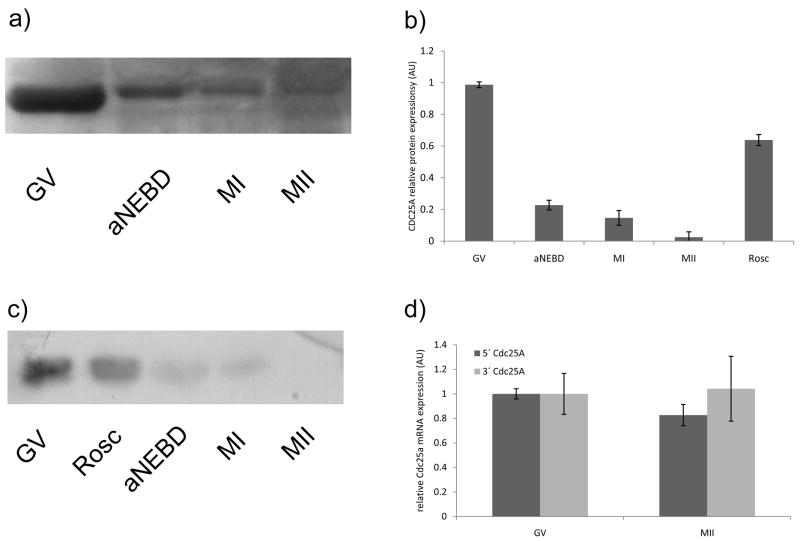
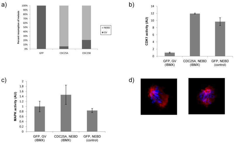
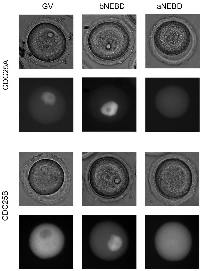
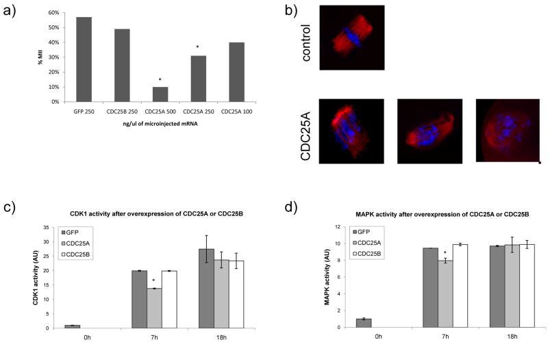
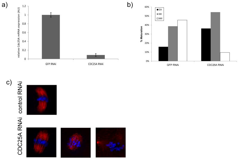
CDK1 is a pivotal regulator of resumption of meiosis and meiotic maturation of oocytes. CDC25A/B/C are dual-specificity phosphatases and activate cyclin-dependent kinases (CDKs). Although CDC25C is not essential for either mitotic or meiotic cell cycle regulation, CDC25B is essential for CDK1 activation during resumption of meiosis. Cdc25a -/- mice are embryonic lethal and therefore a role for CDC25A in meiosis is unknown. We report that activation of CDK1 results in a maturation-associated decrease in the amount of CDC25A protein, but not Cdc25a mRNA, such that little CDC25A is present by metaphase I. In addition, expression of exogenous CDC25A overcomes cAMP-mediated maintenance of meiotic arrest. Microinjection of Gfp-Cdc25a and Gpf-Cdc25b mRNAs constructs reveals that CDC25A is exclusively localized to the nucleus prior to nuclear envelope breakdown (NEBD). In contrast, CDC25B localizes to cytoplasm in GV-intact oocytes and translocates to the nucleus shortly before NEBD. Over-expressing GFP-CDC25A, which compensates for the normal maturation-associated decrease in CDC25A, blocks meiotic maturation at MI. This MI block is characterized by defects in chromosome congression and spindle formation and a transient reduction in both CDK1 and MAPK activities. Lastly, RNAi-mediated reduction of CDC25A results in fewer oocytes resuming meiosis and reaching MII. These data demonstrate that CDC25A behaves differently during female meiosis than during mitosis, and moreover, that CDC25A has a function in resumption of meiosis, MI spindle formation and the MI-MII transition. Thus, both CDC25A and CDC25B are critical for meiotic maturation of oocytes.
Figures
Similar articles
-
CDC25B is required for the metaphase I-metaphase II transition in mouse oocytes.J Cell Sci. 2022 Mar 15;135(6):jcs252924. doi: 10.1242/jcs.252924. Epub 2022 Mar 21. J Cell Sci. 2022. PMID: 35237831
-
Protein kinase A modulates Cdc25B activity during meiotic resumption of mouse oocytes.Dev Dyn. 2008 Dec;237(12):3777-86. doi: 10.1002/dvdy.21799. Dev Dyn. 2008. PMID: 19035343
-
Cdc25b phosphatase is required for resumption of meiosis during oocyte maturation.Nat Genet. 2002 Apr;30(4):446-9. doi: 10.1038/ng856. Epub 2002 Mar 25. Nat Genet. 2002. PMID: 11912493
-
Prophase I arrest and progression to metaphase I in mouse oocytes: comparison of resumption of meiosis and recovery from G2-arrest in somatic cells.Mol Hum Reprod. 2010 Sep;16(9):654-64. doi: 10.1093/molehr/gaq034. Epub 2010 May 7. Mol Hum Reprod. 2010. PMID: 20453035 Free PMC article. Review.
-
Protein kinases and protein phosphatases that regulate meiotic maturation in mouse oocytes.Results Probl Cell Differ. 2011;53:309-41. doi: 10.1007/978-3-642-19065-0_14. Results Probl Cell Differ. 2011. PMID: 21630151 Review.
Cited by
-
The combination of rolipram and cilostamide improved the developmental competence of cloned porcine embryos.Sci Rep. 2023 Apr 7;13(1):5733. doi: 10.1038/s41598-023-32677-3. Sci Rep. 2023. PMID: 37029228 Free PMC article.
-
Investigating SMYD3 role during oocyte maturation in a 3D follicle-enclosed oocyte in vitro model in sheep.Front Cell Dev Biol. 2025 Jun 25;13:1625914. doi: 10.3389/fcell.2025.1625914. eCollection 2025. Front Cell Dev Biol. 2025. PMID: 40636673 Free PMC article.
-
YWHA (14-3-3) protein isoforms and their interactions with CDC25B phosphatase in mouse oogenesis and oocyte maturation.BMC Dev Biol. 2019 Oct 22;19(1):20. doi: 10.1186/s12861-019-0200-1. BMC Dev Biol. 2019. PMID: 31640562 Free PMC article.
-
Insights into molecular features of Venerupis decussata oocytes: a microarray-based study.PLoS One. 2014 Dec 3;9(12):e113925. doi: 10.1371/journal.pone.0113925. eCollection 2014. PLoS One. 2014. PMID: 25470487 Free PMC article.
-
Ser149 is another potential PKA phosphorylation target of Cdc25B in G2/M transition of fertilized mouse eggs.J Biol Chem. 2011 Mar 25;286(12):10356-66. doi: 10.1074/jbc.M110.150524. Epub 2011 Jan 6. J Biol Chem. 2011. PMID: 21212267 Free PMC article.
References
-
- Baldin V, et al. Nuclear localization of CDC25B1 and serine 146 integrity are required for induction of mitosis. J Biol Chem. 2002;277:35176–82. - PubMed
-
- Baldin V, et al. PKB/Akt phosphorylates the CDC25B phosphatase and regulates its intracellular localisation. Biol Cell. 2003;95:547–54. - PubMed
-
- Cazales M, et al. CDC25B phosphorylation by Aurora-A occurs at the G2/M transition and is inhibited by DNA damage. Cell Cycle. 2005;4:1233–8. - PubMed
-
- Davezac N, et al. Regulation of CDC25B phosphatases subcellular localization. Oncogene. 2000;19:2179–85. - PubMed
Publication types
MeSH terms
Substances
Grants and funding
LinkOut - more resources
Full Text Sources
Molecular Biology Databases
Research Materials
Miscellaneous
