. 2008 Jun 5;375(2):611-23.
doi: 10.1016/j.virol.2008.02.025. Epub 2008 Mar 26.

HPV E7 contributes to the telomerase activity of immortalized and tumorigenic cells and augments E6-induced hTERT promoter function

Affiliations

HPV E7 contributes to the telomerase activity of immortalized and tumorigenic cells and augments E6-induced hTERT promoter function

Xuefeng Liu et al. Virology. .

Abstract

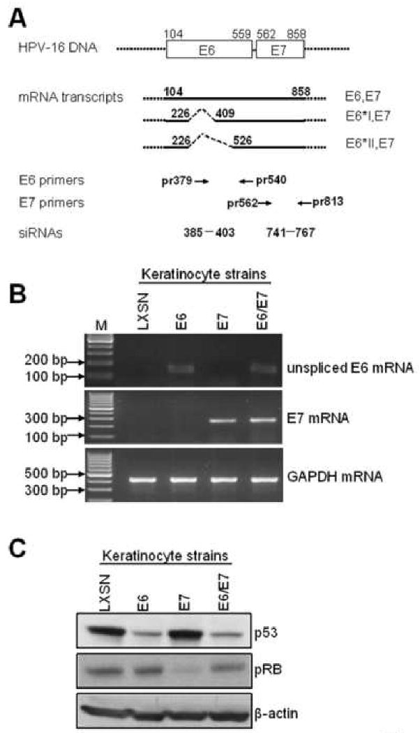
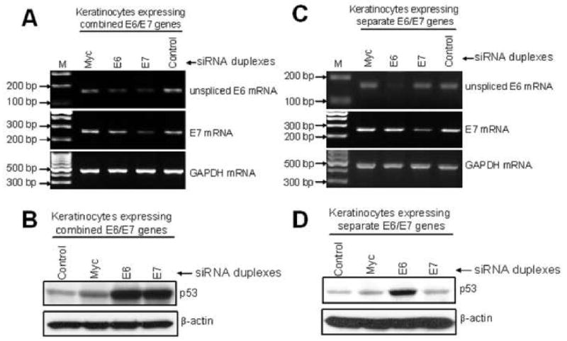
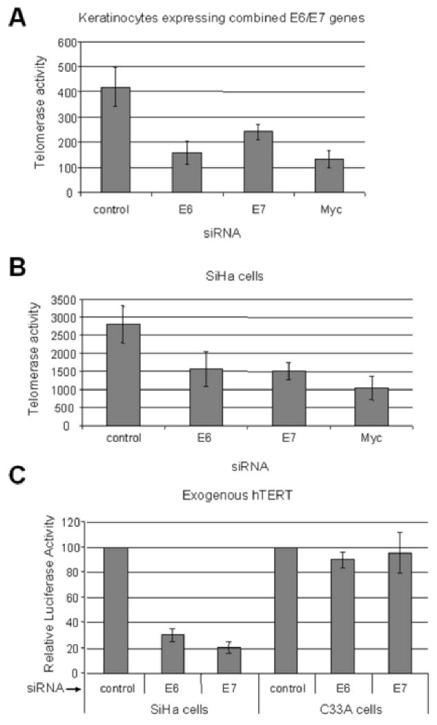
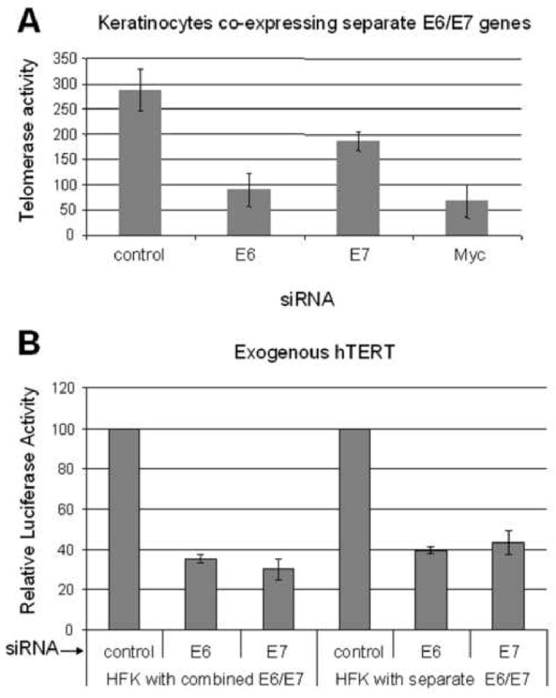
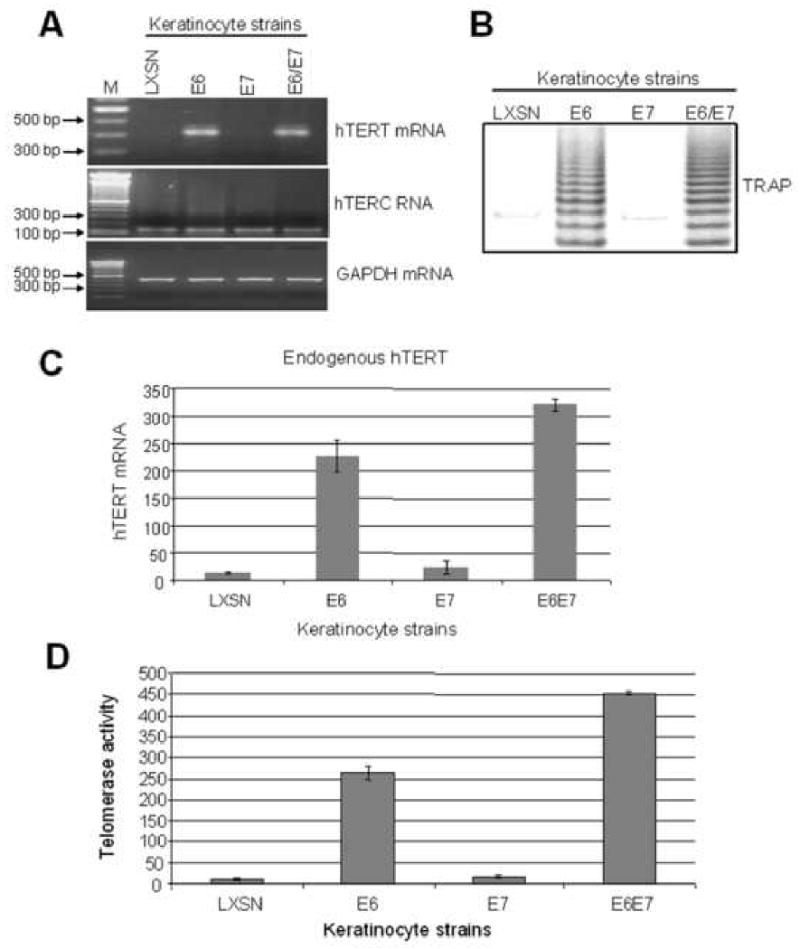
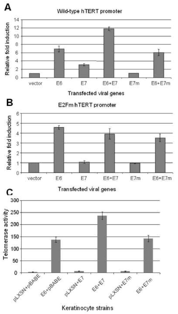
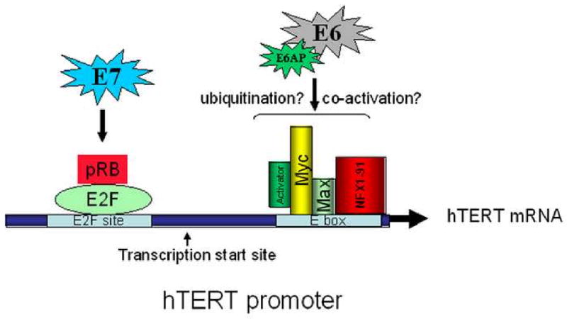
The E6 and E7 proteins of high-risk HPVs are both required for the immortalization of primary human keratinocytes and the maintenance of the malignant phenotype of HPV-positive cancer cell lines. Our previous studies have shown that E6 protein binds Myc protein and that both E6 and Myc associate with and cooperatively activate the hTERT promoter, thereby increasing cellular telomerase activity. In this study, we evaluated the role of E7 in the maintenance and activation of telomerase in immortalized and tumorigenic cells. siRNA knockdown of either E6 or E7 (or both) in HPV-immortalized cells or an HPV-positive cancer cell line reduced hTERT transcription and telomerase activity. Since telomerase was inhibited by E7 siRNA in cells that independently expressed the E6 and E7 genes, our results reveal an independent role for E7 in the maintenance of telomerase activity. However, E7 alone was insufficient to increase endogenous hTERT mRNA or telomerase activity, although it significantly augmented E6-induced hTERT transcription and telomerase activity. To further explore this apparent E7-induced promoter augmentation, we analyzed an exogenous hTERT core promoter in transduced keratinocytes. E7 alone induced the wt hTERT promoter and augmented E6-induced hTERT promoter activity. Mutation of the E2F site in the hTERT promoter abrogated the ability of E7 to induce the hTERT promoter or to enhance the ability of E6 to induce the promoter. Correspondingly, keratinocytes expressing E6 and a mutant E7 (defective for binding pRb pocket proteins) showed lower telomerase activity than cells expressing wt E6 and wt E7. Thus, HPV E7 plays a role in the maintenance of telomerase activity in stable cell lines and augments acute, E6-induced hTERT promoter activity.


Figures

Fig. 1
Fig. 1. Generation and characterization of keratinocyte strains expressing the HPV-16 E6 and E7 genes
(A): Diagram of HPV-16 E6 and E7 mRNA expression and locations of primers and siRNA targets used in this study. The E6 and E7 open reading frames (ORFs) are shown as open boxes, with the nucleotide positions of the first nucleotide of the start codons and the last nucleotide of the stop codons indicated. The dotted lines flanking the open boxes and the heavy lines represent vector sequences. The heavy lines below the open boxes represent the E6/E7 bicistronic transcripts with alternative splicing sites depicted as dotted lines. The numbers above the full length E6E7 transcript are the nucleotide positions of the first nucleotide of the start codon of E6 and the last nucleotide of the stop codon of E7 in the HPV16 genome. The numbers above the E6*IE7 and E6*IIE7 transcripts are the positions of either 5′ or 3′ splicing sites. The primers used in this study are depicted below the transcript lines as arrows and numbers showing the locations of primers in the genome. The siRNA targets are also depicted as dashes and numbers showing the start and end nucleotides of the targets. (B): confirmation of E6, E7 mRNA expression. Primary human foreskin keratinocytes (HFK) were transduced with pLXSN expressing HPV-16 E6, E7, E6E7 or empty vector as described. Total cellular RNA was isolated from the transduced HFK cell strains and RT-PCR was performed with sets of HPV16 unspliced E6 and E7 specific primers described in Material and Methods, GAPDH mRNA was detected as an internal control. PCR products were analyzed with 2% agarose gel. (C): p53 and pRb proteins. To verify the expression of the E6 and E7 proteins, we screened the above stable cell lines by Western blotting for expression of p53 and pRb, respectively. Western blotting for β-actin also was used as a loading control. As anticipated, a decreased p53 protein was observed in E6 and E6E7 expressing cells, and decreased pRb was only observed in E7-expressing cells.
Fig. 2
Fig. 2. siRNA knockdown of E6 and E7 expression in E6E7 transduced keratinocytes
(A) RT-PCR detection of E6 and E7 mRNA (upper and middle panels) and GAPDH mRNA (bottom panel) in keratinocytes expressing the combined E6/E7 genes. The PCR signal for E6 mRNA was reduced in cells treated with siRNA for either E6 or E7. E7 mRNA was decreased in cells treated with siRNA for E7 only. Myc and control siRNA duplexes did not affect the expression of either E6 or E7. The expression of GAPDH mRNA was unaffected by any of these treatments. (B) Western blot detection of p53 protein (upper panel) and β-actin (bottom panel) in HFKs treated with siRNAs. Keratinocytes were treated identically with siRNAs for E6, E7, or Myc as described in (A) but were then analyzed for p53 protein expression by Western blot. p53 protein was increased in cells treated with either E6 or E7 siRNAs, consistent with their postulated inhibition of E6 protein expression. Cells treated with Myc or control siRNA showed lower levels of p53, consistent with continued E6 expression. β-actin protein expression was not altered by any siRNA treatment. (C) RT-PCR detection of E6 and E7 mRNA (upper and middle panels) and GAPDH mRNA (bottom panel) in HFKs that express E6 and E7 from separate vectors. In contrast to experiments with cells expressing the combined E6/E7 genes (panels A-B), cells expressing the E6 and E7 genes from separate vectors showed a specific inhibition by respective siRNAs. E6 mRNA was reduced only when treated with E6 siRNA and E7 mRNA was decreased only in cells treated with E7 siRNA. Myc and control siRNA duplexes had no effect on E6 or E7 expression. The expression of GAPDH mRNA was unaffected by these treatments. (D) Western blot detection of p53 protein (upper panel) and β-actin (bottom panel) in HFKs expressing the E6 and E7 genes from separate vectors. p53 was increased only in cells that were treated with E6 siRNA, indicating specific knockdown of E6 in these cells. These results also verify the results in panel C demonstrating that the E7 siRNA is not inhibiting E6 expression.
Fig. 3
Fig. 3. E6/E7 contribute to the telomerase activity of E6/E7 immortalized HFKs and HPV-positive tumor cells
(A) Telomerase activity inhibition by siRNAs in E6/E7 expressing HFKs. Telomerase activity was measured with a quantitative assay in cells treated with the above siRNAs. Consistent with their ability to inhibit E6, the siRNAs for E6 and E7 decreased telomerase activity by approximately 50%. Myc siRNA was used as a positive control since it has been shown to inhibit cellular telomerase activity. (B) Telomerase activity in SiHa cells after siRNA transfection. Telomerase activity was also reduced in SiHa cancer cells by approximately 50% after treatment with either the E6 or E7 siRNA duplexes. However, since the SiHa cells express E6/E7 coordinately, we cannot conclude an independent role for E7 from these experiments. (C) hTERT promoter activity in tumor cells with siRNA treatment. The wild-type hTERT promoter reporter vector was transfected into the indicated cells together with siRNA for either E6, E7, or control. The value of the promoter activity with control siRNA was set to 100. The hTERT activity was reduced in SiHa cells after treatment with either E6 or E7 specific siRNA by approximately 70% and 80% respectively. Importantly, in cervical cancer cells which lack HPV-16 E6/E7 (C33A), the siRNAs showed no significant inhibition of telomerase activity.
Fig. 4
Fig. 4. E7 contributes to the telomerase activity of E6/E7 immortalized cells
(A) Telomerase activity is dependent upon E7. HFKs expressing the separate E6 and E7 genes were transfected with either the E6 and E7 siRNAs and quantified for telomerase activity as described. Both siRNAs decreased telomerase activity when transfected into recipient cells. Thus, E7 is required for full telomerase activity and its effect on the hTERT promoter is independent of E6. As shown previously in panel C, Myc siRNA also inhibited telomerase activity. (B) hTERT promoter activity is also dependent upon E7. An hTERT promoter reporter construct was transfected into the indicated cells together with siRNA for either E6, E7, or control siRNA. The value of the promoter activity with control siRNA was set to 100. hTERT promoter activity was reduced dramatically in both types of HFKs after treatment with either E6 or E7 specific siRNA. However, only the experiment with HFKs expressing the separate E6 and E7 genes conclusively demonstrates the specific requirement for E7 in maintaining full telomerase activity.
Fig. 5
Fig. 5. E7 augments E6-induced hTERT transcription and telomerase activity
(A) hTERT and hTERC mRNA expression in stable keratinocyte cell lines. hTERT mRNA and hTERC RNA were detected with sets of the primers described in Materials and Methods. Detectable hTERT mRNA was observed only in cells expressing the E6 gene. hTERC RNA was expressed constitutively in all cell strains. (B) Telomerase activity. A qualitative TRAP assay was performed as described in Materials and Methods. A typical DNA ladder (6-base pair difference per band) was generated by cellular telomerase in the cell extract of keratinocytes transduced with the E6 or E6E7 genes. (C) Quantitative RT-PCR detection of hTERT mRNA. A real time quantitative RT-PCR was performed to quantify hTERT mRNA levels shown in panel A. These quantitative data demonstrated reproducible, higher hTERT mRNA levels in E6/E7 expressing cells than in E6 expressing cells. (D) Quantitative TRAP detection of telomerase activity. A quantitative-TRAP assay (Materials and Methods) was also performed to quantify the changes observed in panel B. The E6/E7 cells showed higher telomerase activity than E6 cells, correlating with the hTERT mRNA levels shown in panel C.
Fig. 6
Fig. 6. E7 induces the hTERT promoter through an intact E2F site
(A) Diagram of the hTERT core promoter. Binding sites for transcription factors are indicated as well as the transcription and translation start sites. The sequences and positions of wt and mutated E2F are also depicted. (B) E7 augments E6-induced hTERT promoter activity and this augmentation requires an intact E2F binding site. The wild-type hTERT core promoter (pGL3B-hTERT) was mutated at the E2F binding site using an overlapping PCR protocol described in Materials and Methods. Keratinocytes were transfected with either wt hTERT core promoter (pGL3B-hTERT) or the E2Fm mutant (pGL3B-hTERT-E2Fm) and either E6, E7 or both. The pRL-CMV R. reniformis reporter plasmid was also transfected into the cells to standardize for transfection efficiency. Relative fold activation reflects the normalized luciferase activity induced by E6 and E7 compared to the normalized activity of vector control. The value of pGL3B-hTERT activity with empty vector was set to 1. Error bars show the standard deviation for at least three independent experiments. The mutated E2F binding site in the core hTERT promoter led to an increased basal activity of the promoter and abrogated E7 induction and augmentation of the promoter. Mutation of the E2F binding site did not affect E6 induction of the promoter compared to vector.
Fig. 7
Fig. 7. Augmentation of E6-induced hTERT expression and telomerase activity by E7 requires RB binding
(A) Mutation of the E2F binding site in E7 abolishes the enhancement of hTERT transcription. Wild type and E2Fm promoters (expressing luciferase) were transfected into keratinocytes along with either, E6, E7 or an E7 mutant (E7m), which is unable to bind and degrade pRb. In contrast to wild-type E7, E7m failed to induce the core hTERT promoter and augment E6-induced promoter activity. (B) Neither wt E7 nor E7m induce the hTERT promoter containing an mutated E2F binding site. The mutated hTERT prompter (E2Fm) was transfected into keratinocytes with expression vectors described in panel A. The activity of vector control was set to 1. Both the wt and mutant E7 proteins were unable to induce the mutated hTERT promoter or augment E6-induced promoter activity. (C) Wild-type E7, but not E7m, augments E6-induced telomerase activity in HFKs. Primary HFKs were doubly transduced with two types of retroviruses as described in Materials and Methods. The cell strains were analyzed using a quantitative telomerase assay. Paralleling the results observed with transcription of the hTERT promoter (panel A), we observed that the E7m protein was unable to enhance E6-induced telomerase activity.
Fig. 8
Fig. 8. A working model for regulation of the hTERT promoter by E6 and E7
Results from the current and previous studies are summarized to illustrate the possible mechanism for regulation of the hTERT promoter by the HPV E6 and E7 oncoproteins. A critical regulator of hTERT transcription is the binding of Myc/Max to the promoter E box, as well as the physical interaction of E6/E6AP with this E box site. In addition, the interaction of E7 with the pRb/E2F site is also involved in regulating promoter activity. The transcription start site for hTERT mRNA is indicated.

Similar articles

Cited by

References

    1. Alfandari J, Shnitman Magal S, Jackman A, Schlegel R, Gonen P, Sherman L. HPV16 E6 oncoprotein inhibits apoptosis induced during serum-calcium differentiation of foreskin human keratinocytes. Virology. 1999;257(2):383–96. - PubMed
    1. Alonso MM, Fueyo J, Shay JW, Aldape KD, Jiang H, Lee OH, Johnson DG, Xu J, Kondo Y, Kanzawa T, Kyo S, Bekele BN, Zhou X, Nigro J, McDonald JM, Yung WK, Gomez-Manzano C. Expression of transcription factor E2F1 and telomerase in glioblastomas: mechanistic linkage and prognostic significance. J Natl Cancer Inst. 2005;97(21):1589–600. - PubMed
    1. Alonso MM, Fueyo J, Yung WK, Gomez-Manzano C. E2F1 and telomerase: alliance in the dark side. Cell Cycle. 2006;5(9):930–5. - PubMed
    1. Amati B. Myc degradation: dancing with ubiquitin ligases. Proc Natl Acad Sci U S A. 2004;101(24):8843–4. - PMC - PubMed
    1. Androphy EJ, Hubbert NL, Schiller JT, Lowy DR. Identification of the HPV-16 E6 protein from transformed mouse cells and human cervical carcinoma cell lines. Embo J. 1987;6(4):989–92. - PMC - PubMed

Publication types