. 2008 Jun 5;375(2):624-36.
doi: 10.1016/j.virol.2008.02.023. Epub 2008 Mar 26.

An Amsacta moorei entomopoxvirus ortholog of the poly(A) polymerase small subunit exhibits methyltransferase activity and is non-essential for virus growth

Affiliations

An Amsacta moorei entomopoxvirus ortholog of the poly(A) polymerase small subunit exhibits methyltransferase activity and is non-essential for virus growth

Marie N Becker et al. Virology. .

Abstract

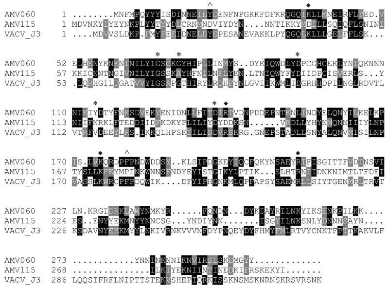
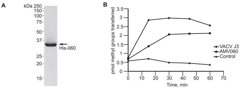
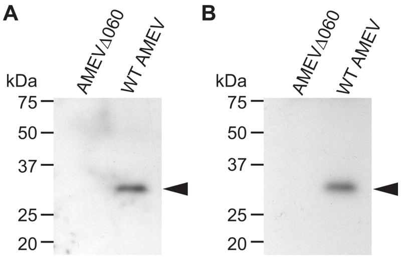
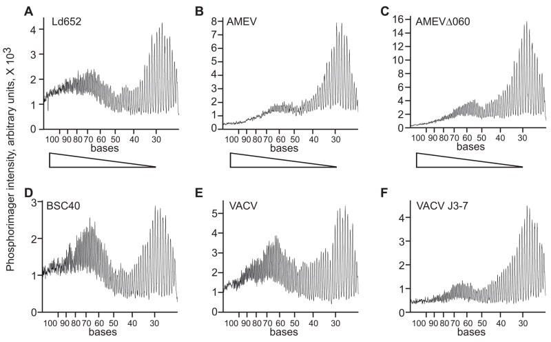
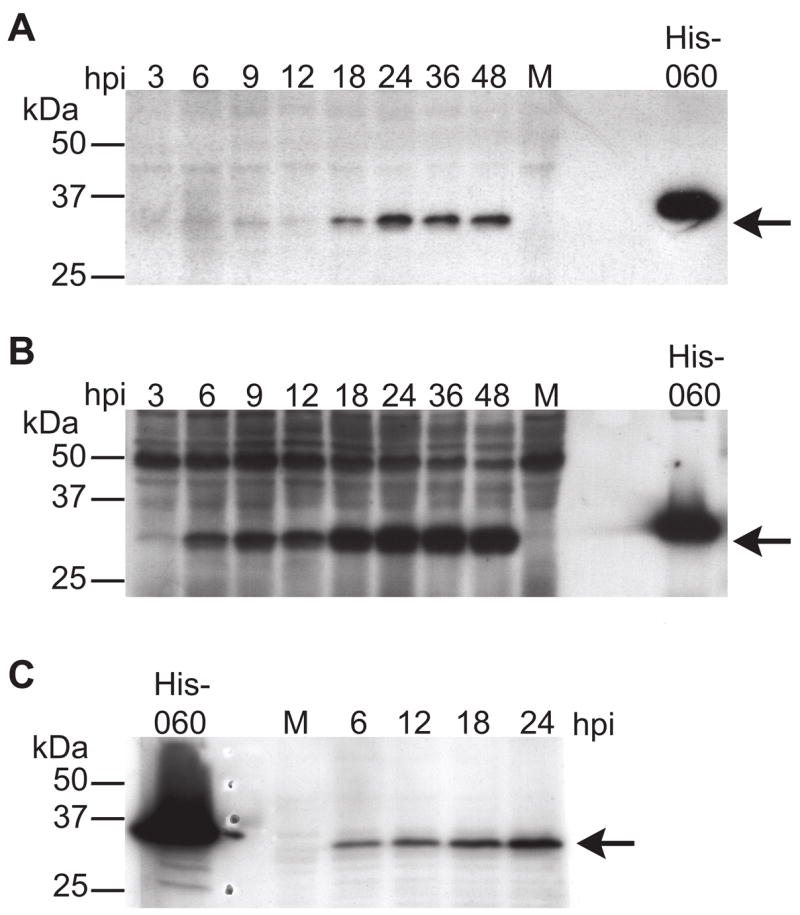
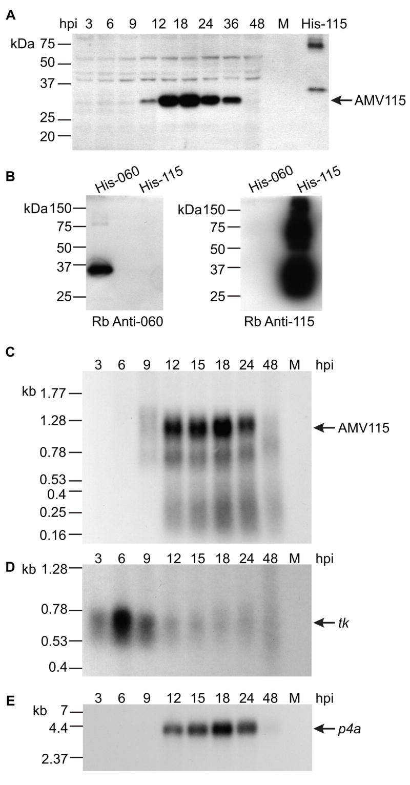
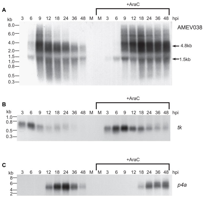
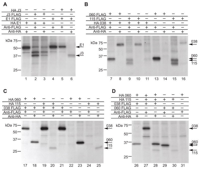
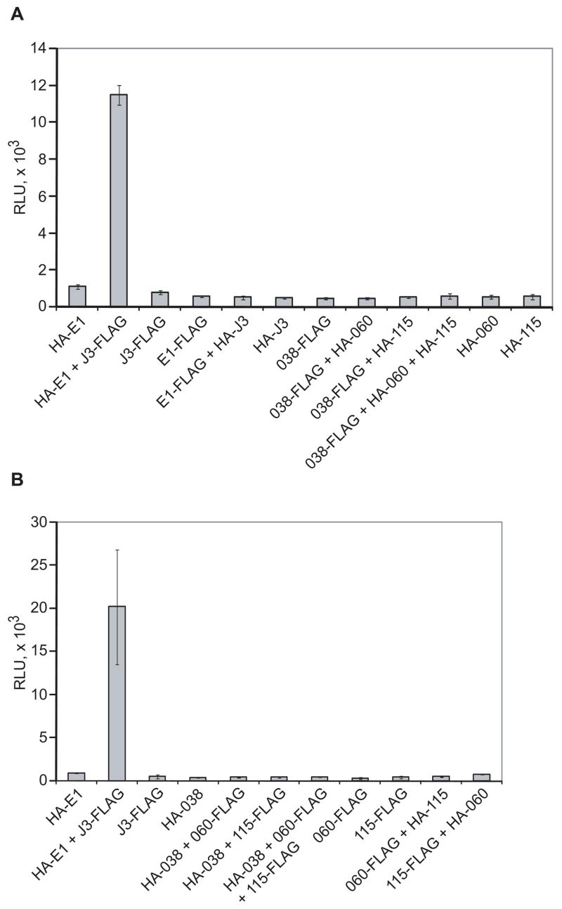
Unlike the heterodimeric poly(A) polymerase (PAP) of vaccinia virus (VACV), the PAP from the Amsacta moorei entomopoxvirus, AMEV, is potentially derived from three subunits: a single large and two small subunits (AMV060 and AMV115). The VACV small subunit serves as a 2'-O-methyltransferase, a processivity factor for mRNA polyadenylation, and a transcription elongation factor. We wished to determine the structure-function relationships of the three putative AMEV PAP subunits. We show that AMV060 is expressed as an early gene persisting throughout infection, whereas AMV115 is expressed late. We demonstrate that AMV060 exhibits 2'-O-methyltransferase activity but the gene is not essential for virus growth. Absence of the AMV060 protein has no effect on the length of the poly(A) tails present in mRNA. No physical association was found between any of the putative AMEV PAP subunits. We therefore propose that mRNA polyadenylation does not require interactions between these three proteins.


Figures

Figure 1
Figure 1
Alignment of AMEV and VACV small PAP subunits. Critical amino acids with known functions in the VACV J3 subunit are designated thus: residues involved in cap binding are indicated by ^; those necessary for SAM interactions by *; and residues for methyltransferase function by ♦. Alignment constructed with AlignX program (Invitrogen) which uses a Clustal W algorithim. Identical residues have black backgrounds. Similar residues are gray.
Figure 2
Figure 2
Purification of His-060 protein and methyltransferase activity assay. A. Coomassie stained gel of His tagged AMV060 protein purified via metal chelation and cation exchange chromatography. B. Methyltransferase assay. The incorporation of 3H methyl groups into BMV genomic DNA at 37° by 2μg of either His-060 or VACV His J3 protein indicating 2′-O-methyltransferase activity.
Figure 3
Figure 3
Deletion of the AMV060 gene in AMEV. Immunoblots developed with either A) the chicken anti-060 antisera or B) rabbit anti-060 peptide antisera. The results indicate an absence of the AMV060 protein from AMEVΔ060. Arrowheads indicate the position of the AMV060 protein.
Figure 4
Figure 4
Comparative growth curves for AMEV and AMEVΔ060. A) Growth curves at low multiplicity (MOI 0.01), and B) at high multiplicity (MOI = 10). Virus yields at various time points were determined via plaque assay. Duplicate samples were titered. Error bars indicate the standard deviation.
Figure 5
Figure 5
Analysis of poly(A) tail length. The length of poly(A) tails on mRNA was analyzed from total RNA harvested from cells infected at an MOI=10 at 18hpi. After radiolabeling and RNase digestion, the poly(A) tails which remain were analyzed on a polyacrylamide sequencing gel and visualized on a phosphorimager screen. The length of the poly(A) tails was measured with a Storm phosphorimager and the resultant graphs presented here. The intensity of the bands is graphed in arbitrary units on the ordinate and the scale varies for each sample. The size in bases is graphed on the abscissa decreasing from left to right. RNA from each of the following conditions is shown: A. uninfected Ld652 cells; B. wild type AMEV infected Ld652 cells; C. AMEVΔ060 infected Ld652 cells. Bottom row: D. uninfected BSC40 cells; E. wild type VACV infected BSC40 cells and F. mutant VACV J3-7 infected BSC40 cells.
Figure 6
Figure 6
Time course of AMV060 protein expression. Protein samples were harvested after infection at the times indicated and analyzed on immunoblots with the following antibodies: A. Rabbit anti-060 peptide antibody. B. Chicken anti-060 antibody. C. Polyclonal mouse anti-060 antibody. Arrows indicate wild type 060, mock infected cells are denoted as M, and purified His-060 protein is so designated. The His-060 protein migrates slower than the wild type protein due to the presence of the His tag. In all blots, M represents mock infected cells.
Figure 7
Figure 7
Time course of AMV115 expression. A. Immunoblot with rabbit anti-115 peptide antibody. Protein samples were harvested at the times indicated. M indicates mock infected cells. Partially purified His-115 is shown as a control. The addition of the His-tag appears to lead to a slight retardation in the mobility of the protein compared to the untagged protein. B. Blots containing 0.5 μg each of purified His-060 and His-115 protein detected with either rabbit anti-060 or anti-115 peptide antibodies indicate no cross reactions by these antibodies to the other protein. C. Northern blot probed with an antisense oligonucleotide to AMV115. The RNA samples were harvested from infected Ld652 cells at the times indicated and each lane contains 3μg total RNA. Blots probed with antisense oligonucleotides for either the D. early (tk) and E. late (p4a) genes are shown below (tk and p4a blots taken from Becker et al., 2004).
Figure 8
Figure 8
Northern analysis of AMV038 expression in cells infected in the presence or absence of AraC. All blots contain 3μg of total RNA per lane. RNA was harvested from Ld652 infected with WT AMEV at the time points indicated. A. RNA probed with an AMV038 riboprobe. The left portion of the blot shows AMV038 profile in the absence of AraC. The right portion is RNA harvested from infected cells grown in the presence of 250μg/ml AraC. Control Northern blots for B. the early gene, tk, and C. the late gene, p4a for comparative purposes.
Figure 9
Figure 9
Co-immunoprecipitations of poly(A) polymerase subunits. Radiolabeled protein extracts were derived from BSR-T7 cells that were transfected with plasmids which express the epitope tagged PAP subunits. Extracts were incubated with the indicated antibody and then immunoprecipitated with protein A sepharose. Precipitated proteins were visualized by autoradiography after SDS-PAGE. A. Examination of interactions between VACV subunits E1 and J3 subunits. B. Examination of interactions between FLAG tagged individual small AMEV subunits and the HA tagged large subunit. AMV115 FLAG appears to be expressed as a doublet, lanes 9 and 15. C. Examination of interactions between HA tagged individual small AMEV subunits and the FLAG tagged large subunit. D. Examination of potential interactions between the two small subunits or all three AMEV PAP subunits. The double arrow for 060 indicates the small difference in size between HA-060 and 060-FLAG.
Figure 10
Figure 10
Alpha Screen analysis of potential protein-protein interactions. Protein extracts were made from BSR-T7 cells that were transfected overnight with plasmids designed to express epitope-tagged proteins under control of a bacteriophage T7 promoter. Biotinylated anti-HA antibody, streptavidin donor beads and anti-FLAG receptor beads were incubated with the extracts and light emission was quantified with an EnVision plate reader. Levels of light emission are graphed in relative light units (RLU). Panels A and B represent experiments performed at different times, note the different scales. n=2 for both panels and the average value is presented.

Similar articles

Cited by

References

    1. Afonso CL, Tulman ER, Lu Z, Oma E, Kutish GF, Rock DL. The genome of Melanoplus sanguinipes Entomopoxvirus. Journal of Virology. 1999;73:533–552. - PMC - PubMed
    1. Barbosa E, Moss B. mRNA(nucleoside-2′-)-methyltransferase from vaccinia virus. Purification and physical properties. J Biol Chem. 1978;253:7692–7697. - PubMed
    1. Bawden AL, Glassberg KJ, Diggans J, Shaw R, Farmerie W, Moyer RW. Complete genomic sequence of the Amsacta moorei entomopoxvirus: analysis and comparison with other poxviruses. Virology. 2000;274:120–139. - PubMed
    1. Becker MN, Greenleaf WB, Ostrov DA, Moyer RW. Amsacta moorei entomopoxvirus expresses an active superoxide dismutase. J Virol. 2004;78:10265–10275. - PMC - PubMed
    1. Becker MN, Moyer RW. Subfamily Entomopoxvirinae. In: Mercer A, Schmidt A, Weber O, editors. Poxviruses. Birkhäuser Publishing; Basel: 2007.

Publication types