

A novel serine phosphorylation site detected in the N-terminal domain of estrogen receptor isolated from human breast cancer cells
- PMID: 18367407
- PMCID: PMC7456516
- DOI: 10.1016/j.jasms.2008.02.008
A novel serine phosphorylation site detected in the N-terminal domain of estrogen receptor isolated from human breast cancer cells
Abstract
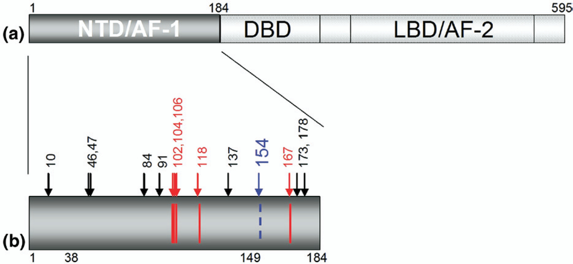
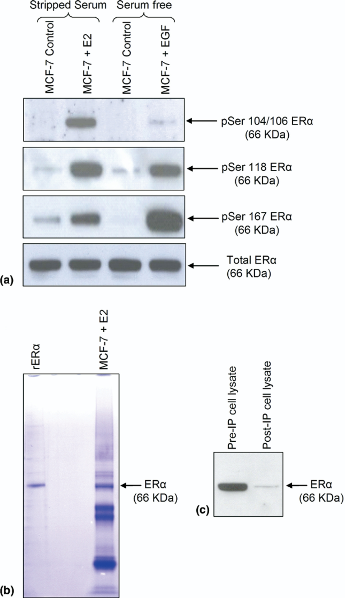
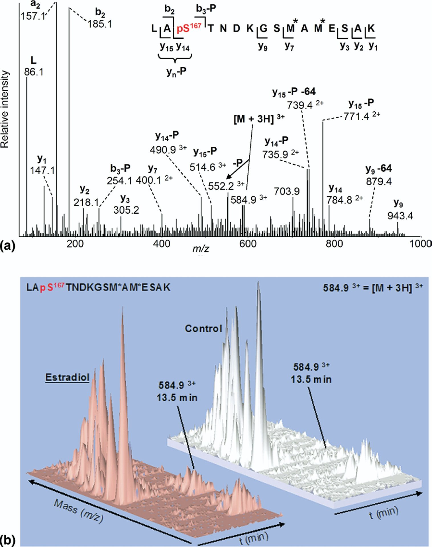
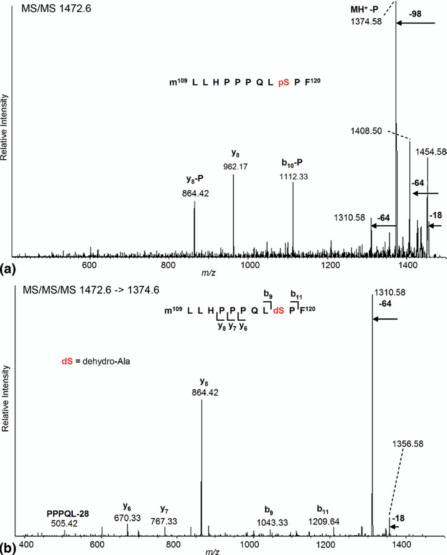
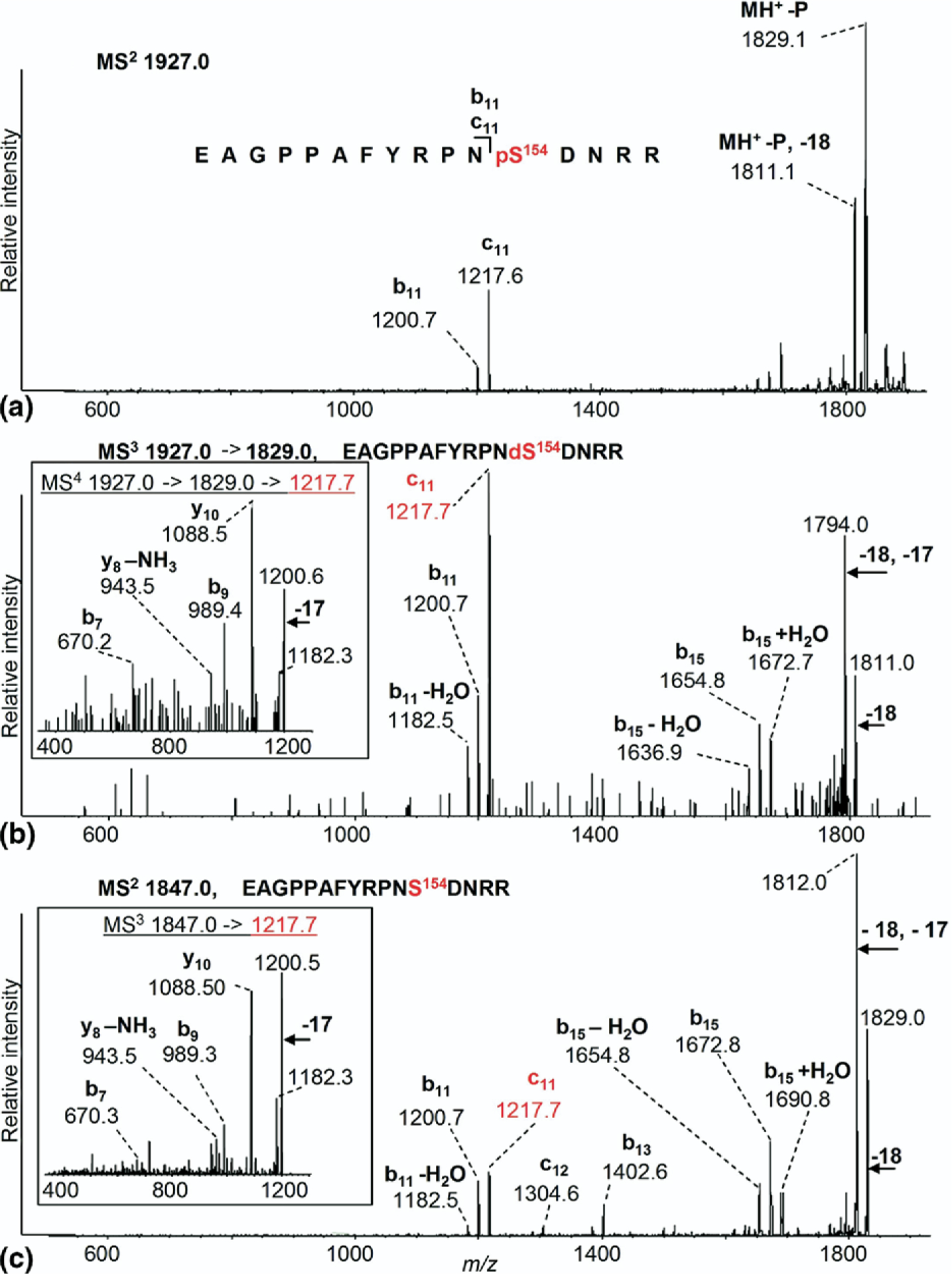
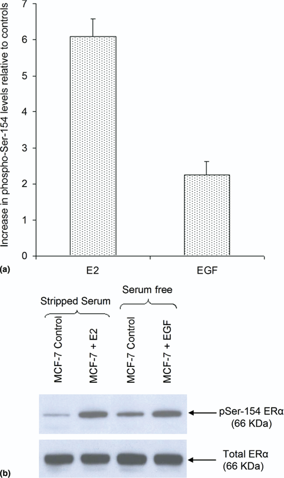
Activated estrogen receptor (ERalpha) plays a critical role in breast cancer development and is a major target for drug treatment. Serine phosphorylation within the N-terminal domain (NTD) contributes to ERalpha activation and may also cause drug resistance. Previous biochemical identification of phosphorylated ERalpha residues was limited to protein artificially overexpressed in transfected cell lines. We report mass spectrometric methods that have allowed the identification of a new site within the NTD of ERalpha isolated from cultured human breast cancer cells. Immunoprecipitation, trypsin digestion, and analysis by nano-LC-ESI-MS/MS (Q-STAR, MDS Sciex) and vMALDI-MS(n) (Finnigan LTQ, Thermo-Electron) identified peptides containing 8 of 14 serine residues within the NTD, one being partially phosphorylated Ser-167, known but not previously reported by MS. Chymotrypsin digestion revealed other known sites at Ser-102/104/106 and 118. Tandem methods developed for the peptide containing Ser-118 and the use of hypothesis-driven experiments--i.e., the assumption that an intact phosphopeptide showing no molecular ion might yield fragment ions including loss of phosphoric acid in vMALDI-MS/MS--allowed the identification of a novel site at Ser-154. Quantitation by selected reaction monitoring demonstrated 6-fold and 2.5-fold increases in Ser-154 phosphorylation in estradiol- and EGF-treated cells, respectively, compared to controls, confirmed by immunoblotting with a novel rabbit polyclonal antibody. Thus, the protein isolation and MS strategies described here can facilitate discovery of novel phosphorylation sites within low abundance, clinically important cancer targets like ERalpha, and may thereby contribute to our understanding of the role of phosphorylation in the development of breast cancer.
Conflict of interest statement
The authors declare no conflict of interest.
Figures
References
-
- Joel PB; Traish AM; Lannigan DA Estradiol-Induced Phosphorylation of Serine 118 in the Estrogen Receptor Is Independent of p42/p44 Mitogen-Activated Protein Kinase. J. Biol. Chem 1998, 273, 13317–13323. - PubMed
-
- Likhite VS; Stossi F; Kim K; Katzenellenbogen BS; Katzenellenbogen JA Kinase-Specific Phosphorylation of the Estrogen Receptor Changes Receptor Interactions with Ligand, Deoxyribonucleic Acid, and Coregulators Associated with Alterations in Estrogen and Tamoxifen Activity. Mol. Endocrinol 2006, 20, 3120–3132. - PubMed
-
- McInerney EM; Katzenellenbogen BS Different Regions in Activation Function-1 of the Human Estrogen Receptor Required for Antiestrogen- and Estradiol-dependent Transcription Activation. J. Biol. Chem 1996, 271, 24172–24178. - PubMed
-
- Jensen EV; Jordan VC The Estrogen Receptor: A Model for Molecular Medicine. Clin. Cancer Res 2003, 9, 1980–1989. - PubMed
-
- Shiau AK; Barstad D; Loria PM; Cheng L; Kushner PJ; Agard DA; Greene GL The Structural Basis of Estrogen Receptor/ Coactivator Recognition and the Antagonism of This Interaction by Tamoxifen. Cell 1998, 95, 927–937. - PubMed
Publication types
MeSH terms
Substances
Grants and funding
LinkOut - more resources
Full Text Sources
Other Literature Sources
Medical
Molecular Biology Databases
Research Materials
Miscellaneous
