. 2008 Nov;12(8):1078-89.
doi: 10.1016/j.ejpain.2008.02.002. Epub 2008 Mar 25.

Brain activity associated with slow temporal summation of C-fiber evoked pain in fibromyalgia patients and healthy controls

Affiliations

Brain activity associated with slow temporal summation of C-fiber evoked pain in fibromyalgia patients and healthy controls

Roland Staud et al. Eur J Pain. 2008 Nov.

Abstract

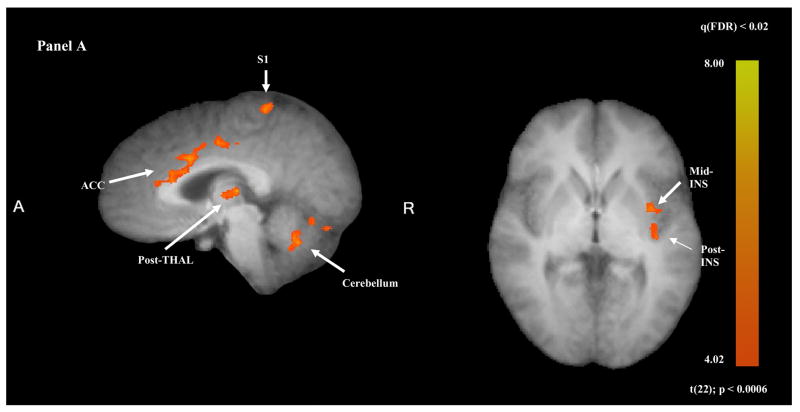
Temporal summation of "second pain" (TSSP) is the result of C-fiber-evoked responses of dorsal-horn neurons, termed "windup". This phenomenon is dependent on stimulus frequency (0.33 Hz) and relevant for central sensitization as well as chronic pain. Whereas, our previous functional magnetic resonance imaging (fMRI) study characterized neural correlates of TSSP in 11 healthy volunteers, the present study was designed to compare brain responses associated with TSSP across these healthy participants and 13 fibromyalgia (FM) patients. Volume-of-interest analysis was used to assess TSSP-related brain activation. All participants underwent fMRI-scanning during repetitive heat pulses at 0.33 Hz and 0.17 Hz to the right foot. Stimulus intensities were adjusted to each individual's heat sensitivity to achieve comparable TSSP-ratings of moderate pain in all subjects. Experimental pain ratings showed robust TSSP during 0.33 Hz but not 0.17 Hz stimuli. When stimulus strength was adjusted to induce equivalent levels of TSSP, no differences in activation of pain-related brain regions occurred across NC and FM groups. Subsequently, the fMRI-data of both groups were combined to increase the power of our statistical comparisons. fMRI-statistical maps identified several brain regions with stimulus and frequency dependent activation consistent with TSSP, including ipsilateral and contralateral thalamus, medial thalamus, S1, bilateral S2, mid- and posterior insula, rostral and mid-anterior cingulate cortex. However, the stimulus temperatures necessary to evoke equivalent levels of TSSP and corresponding brain activity were less in FM patients. These results suggest that enhanced neural mechanisms of TSSP in FM are reflected at all pain related brain areas, including posterior thalamus, and are not the result of selective enhancement at cortical levels.


Figures

Figure 1
Figure 1
Repetitive thermal stimuli at 0.33 Hz and 0.17 Hz were used to image TSSP-related brain activation. Previous psychophysical studies have shown that stimulus frequencies of ≥ 0.33 Hz resulted in robust TSSP whereas frequencies of < 0.33 Hz were only minimally effective. During scanning runs, two and six heat stimuli at 0.33 Hz and 0.17 Hz were applied to the right plantar surface, respectively. We previously used this format of stimulation for the NC subjects (Staud et al. 2007a). To remain consistent, the same paradigm was used for the FM subjects in the current study. Each run lasted for 88 sec and was comprised of discarded acquisitions/baseline, repetitive thermal stimuli at 0.17 Hz or 0.33 Hz, and a 30 sec follow-up period. Although the four different trains used during scanning runs varied in the timing of the first stimulus, the last stimulus of each train always occurred at the same time point of a run. Duration of discarded acquisitions/baselines is indicated by hatched lines; stimuli at 0.33 Hz or 0.17 Hz are shown by solid black lines; the 30 sec follow-up periods are represented by solid grey lines.
Figure 2
Figure 2
Average ratings (SD) of maximal experimental pain during TSSP trials at 0.17 Hz and 0.33 Hz in NC and FM subjects. All subjects underwent eight repetitions of two pulse or six pulse trains at either frequency in the scanner. All experimental pain ratings were obtained at the end of each scanning run using the NPS. The experimental stimuli were adjusted outside the scanner to each individual’s heat sensitivity to obtain TSSP ratings of 45 ± 10 NPS units after 6 stimuli at 0.33 Hz. inside the scanner, however, the mean
Figure 3
Figure 3
Brain activity related to TSSP stimuli in sagittal, axial, and coronal sections (Panel A and B). Data of NC and FM subjects were combined for this analysis. The fMRI images depict the difference in brain activation between the sixth (last) stimulus at 0.33 Hz and all other stimulus conditions (including the 2-pulse trains at 0.33 Hz and 2- and 6-pulse trains at 0.08 Hz). The brain activity in all a-priori selected VOIs was significantly greater after six pulses at 0.33 Hz compared to all other conditions at, including S1, ACC, THAL, INS, and cerebellum (p < .006). Statistical maps are overlaid on structural MRIs averaged across all study participants. (THAL: thalamus; S1: somato-sensory cortex 1; ACC: anterior cingulate cortex; INS: insula).
Figure 3
Figure 3
Brain activity related to TSSP stimuli in sagittal, axial, and coronal sections (Panel A and B). Data of NC and FM subjects were combined for this analysis. The fMRI images depict the difference in brain activation between the sixth (last) stimulus at 0.33 Hz and all other stimulus conditions (including the 2-pulse trains at 0.33 Hz and 2- and 6-pulse trains at 0.08 Hz). The brain activity in all a-priori selected VOIs was significantly greater after six pulses at 0.33 Hz compared to all other conditions at, including S1, ACC, THAL, INS, and cerebellum (p < .006). Statistical maps are overlaid on structural MRIs averaged across all study participants. (THAL: thalamus; S1: somato-sensory cortex 1; ACC: anterior cingulate cortex; INS: insula).
Figure 4
Figure 4
Representative samples of BOLD signal time courses related to six TSSP stimuli at 0.33 Hz (black lines) and 0.17 Hz (grey lines) of NC and FM subjects. VOIs: left rostral ACC; left THAL. Arrows indicate the duration of six heat stimuli at 0.33 Hz (black arrow) and 0.17 Hz (grey arrow). Residual brain activation after 0.33 Hz 6-pulse trains took more than twice as long to resolve as expected. In contrast residual brain activity after 0.17 Hz 6-pulse trains resolved within the expected time period. Similar BOLD time courses were observed at all VOIs. Signal intensity time courses were averaged across all significant voxels within statistically-identified clusters. TSSP: temporal summation of second pain; THAL: thalamus; BOLD: blood oxygen level dependent.
Figure 5
Figure 5
Representative samples of BOLD signal time courses related to six TSSP stimuli at 0.33 Hz (black lines) and 0.17 Hz (grey lines) of all subjects (NC and FM) combined. VOIs: left rostral ACC; left THAL. Arrows indicate the duration of six heat stimuli at 0.33 Hz (black arrow) and 0.17 Hz (grey arrow). Brain activity was most pronounced during six heat stimuli at 0.33 Hz. Similar BOLD time courses were observed at all VOIs. Signal intensity time courses were averaged across all significant voxels within statistically-identified clusters. TSSP: temporal summation of second pain; THAL: thalamus; BOLD: blood oxygen level dependent

References

    1. Al Allaf AW, Khan F, Moreland J, Belch JJF, Pullar T. Investigation of cutaneous microvascular activity and flare response in patients with fibromyalgia syndrome. Rheumatology. 2001;40(10):1097–1101. - PubMed
    1. Beck AT, Beamesderfer A. Assessment of depression: the depression inventory. In: Pichot P, editor. Psychological measurements in psychopharmacology. Basel: Karger; 1974. pp. 151–169. - PubMed
    1. Beitel RE, Dubner R. Response of unmyelinated (C) polymodal nociceptors to thermal stimuli applied to monkey’s face. J Neurophysiol. 1976;39(6):1160–1175. - PubMed
    1. Cook DB, Lange G, Ciccone DS, Liu WC, Steffener J, Natelson BH. Functional imaging of pain in patients with primary fibromyalgia. J Rheumatol. 2004;31(2):364–378. - PubMed
    1. Desmeules JA, Cedraschi C, Rapiti E, Baumgartner E, Finckh A, Cohen P, Dayer P, Vischer TL. Neurophysiologic evidence for a central sensitization in patients with fibromyalgia. Arthritis Rheum. 2003;48(5):1420–1429. - PubMed

Publication types

MeSH terms