. 2008 Jun 6;283(23):16135-46.
doi: 10.1074/jbc.M710323200. Epub 2008 Mar 26.

Crystal structure of human spermine synthase: implications of substrate binding and catalytic mechanism

Affiliations

Crystal structure of human spermine synthase: implications of substrate binding and catalytic mechanism

Hong Wu et al. J Biol Chem. .

Abstract

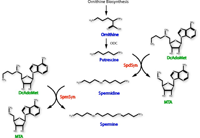
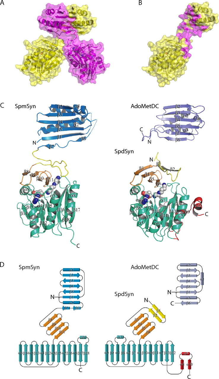
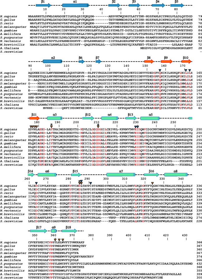
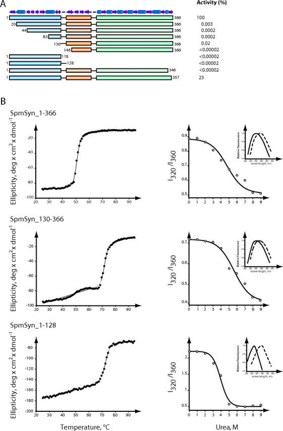
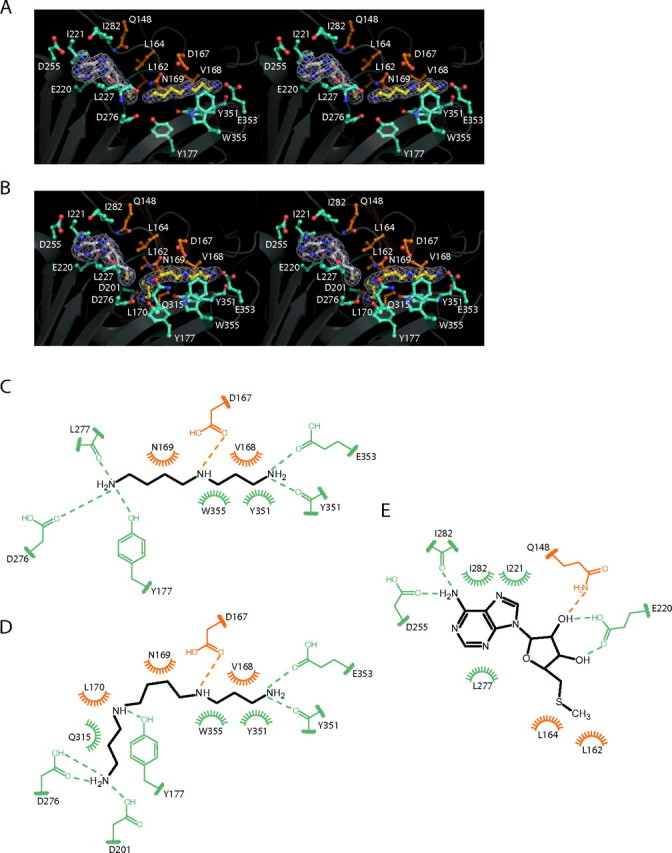
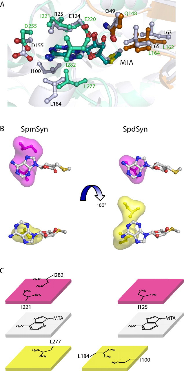
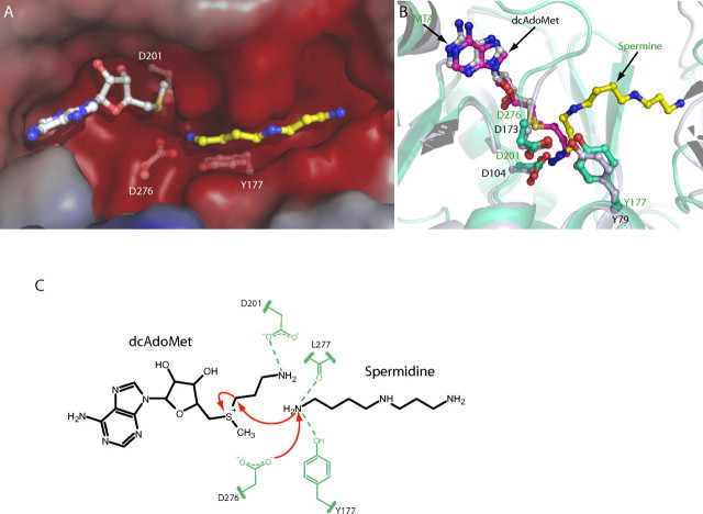
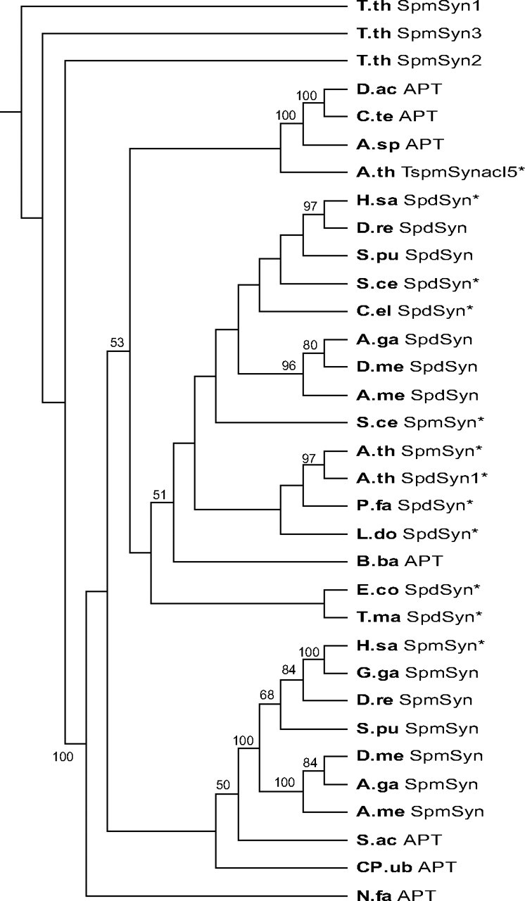
The crystal structures of two ternary complexes of human spermine synthase (EC 2.5.1.22), one with 5'-methylthioadenosine and spermidine and the other with 5'-methylthioadenosine and spermine, have been solved. They show that the enzyme is a dimer of two identical subunits. Each monomer has three domains: a C-terminal domain, which contains the active site and is similar in structure to spermidine synthase; a central domain made up of four beta-strands; and an N-terminal domain with remarkable structural similarity to S-adenosylmethionine decarboxylase, the enzyme that forms the aminopropyl donor substrate. Dimerization occurs mainly through interactions between the N-terminal domains. Deletion of the N-terminal domain led to a complete loss of spermine synthase activity, suggesting that dimerization may be required for activity. The structures provide an outline of the active site and a plausible model for catalysis. The active site is similar to those of spermidine synthases but has a larger substrate-binding pocket able to accommodate longer substrates. Two residues (Asp(201) and Asp(276)) that are conserved in aminopropyltransferases appear to play a key part in the catalytic mechanism, and this role was supported by the results of site-directed mutagenesis. The spermine synthase.5'-methylthioadenosine structure provides a plausible explanation for the potent inhibition of the reaction by this product and the stronger inhibition of spermine synthase compared with spermidine synthase. An analysis to trace possible evolutionary origins of spermine synthase is also described.


Figures

FIGURE 1.
FIGURE 1.
General pathway for polyamine synthesis. ODC, ornithine decarboxylase.
FIGURE 2.
FIGURE 2.
Structure of SpmSyn and structural homology to AdoMetDC and SpdSyn. A, dimer of SpmSyn. The two chains are in yellow and magenta. The enzyme is shown in schematic and surface representations; the bound molecules of SPD and MTA are shown in sphere representation, with carbon atoms in gray. B, monomer of SpmSyn with the dimerization interface in magenta. C, structures of the SpmSyn monomer, the SpdSyn monomer (Protein Data Bank code 2O06), and AdoMetDC (Protein Data Bank code 1TLU) shown in schematic representation. The N-terminal, central, and C-terminal domains in SpmSyn are in blue, orange, and greencyan, respectively. The loop connecting the N-terminal and central domains is in yellow. AdoMetDC is in purple. The SpdSyn domain color coding is the same as for SpmSyn, except for the first two β-strands, which structurally align with long interdomain loop of SpmSyn, are in yellow, and three C-terminal helices, which are absent in SpmSyn, are in red. SPD and MTA bound to SpmSyn and SpdSyn are shown in sphere representation, with carbon atoms in gray. The figures were generated using the program PyMOL (DeLano Scientific, San Carlos, CA). D, topology diagram for SpmSyn, SpdSyn, and AdoMetDC. All of the secondary structure elements are labeled. All of the domains in each molecule are colored as described for C.
FIGURE 3.
FIGURE 3.
Multiple sequence alignment of representative members of SpmSyn family. Identical residues are colored in red in the alignment. Secondary structure elements of human SpmSyn were assigned by the PROCHECK program (58) and are shown above the sequences and labeled: the helices are shown as cylinders, and the strands are shown as arrows. The three domains in human SpmSyn are colored in blue, orange, and greencyan. Catalytic residues are labeled with asterisks; the residues interacting with SPD and SPM are labeled with orange arrowheads; and the residues involved in MTA binding are labeled with blue arrowheads. The alignment was generated using ClustalW (59) and assisted with hand fittings. The sequences shown are from human (Homo sapiens; NP_004586), chicken (Gallus gallus; NP_001025974), zebrafish (Danio rerio; NP_571831), fruit fly (Drosophila melanogaster; NP_729798), mosquito (Anopheles gambiae; XP_315341), bee (Apis mellifera; XP_393567), sea urchin (S. purpuratus; XP_789223, 25 amino acids truncated from the C terminus), sea anemone (N. vectensis; XP_001636780, N terminus extended 50 amino acids using expressed sequence tag DV091768), M. brevicollis (Joint Genome Institute protein ID 30201), A. thaliana (NP_568785), and Saccharomyces cerevisiae (AAC19368).
FIGURE 4.
FIGURE 4.
Enzymatic activity and tertiary structure of SpmSyn fragments. A, activity of each truncated protein shown as the percentage of the activity of the full-length enzyme. The three domains are colored in blue, orange, and greencyan. The secondary structure elements of SpmSyn are shown as cylinders (for helices) and arrows (for strands) on the top.B, analysis of the tertiary structure of full-length SpmSyn and its domains. The left panels show the thermo-melting profiles of full-length SpmSyn and its fragments. The ellipticity was measured at 220 nm. The right panels show the dependence of fluorescence spectra (I320/I360) of human SpmSyn and its fragments in the presence of different concentrations of urea. Insets show the fluorescence spectra in native buffer (20 mm Tris-HCl (pH 7.5) and 150 mm NaCl; solid lines) and in the presence of 9 m urea (dashed lines). The boundary of each construct is labeled on top of the profile. deg, degrees.
FIGURE 5.
FIGURE 5.
Active site of SpmSyn. A and B, stereo views of the active sites of SpmSyn in the SpmSyn·MTA·SPD and SpmSyn·MTA·SPM ternary complexes, respectively. The enzyme is shown in schematic representation. The MTA, SPD, and SPM molecules are shown in stick-and-ball representation, with carbon atoms in MTA in gray and carbon atoms in SPD and SPM in yellow. The 2mFo - DFc map of each ligand is shown in mesh (contoured to 1.0σ). All of the residues interacting with the ligands are shown in stick-and-ball representation. The carbon atoms of residues from the central domain of SpmSyn are in orange, whereas the carbon atoms of residues from the C-terminal domain are in greencyan. C-E, schematic illustrations of interactions between the activesite residues and SPD, SPM, and MTA, respectively. Dashed lines represent hydrogen bonds, and arcs represent hydrophobic interactions. The residues from the central and C-terminal domains are in orange and greencyan.
FIGURE 6.
FIGURE 6.
Structural basis for inhibition of SpmSyn by MTA. A, superimposition of MTA-binding sites in SpmSyn and SpdSyn. The structures of SpmSyn (Protein Data Bank code 3C6K) shown in greencyan and SpdSyn (Protein Data Bank code 2O0L) shown in gray are shown in schematic representation and superimposed. MTA and the residues interacting with MTA in both structures are shown in stick-and-ball representation, and the carbon atoms are in the same color as the corresponding structure, except for the three residues from the central domain of SpmSyn, which are in orange. B and C, illustrations of the hydrophobic surfaces interacting with the adenine ring in MTA. The two surfaces stacking with the adenine ring are in magenta and yellow. MTA is shown in stick-and-ball representation, with carbon atoms in gray.
FIGURE 7.
FIGURE 7.
dcAdoMet-binding site of SpmSyn and illustration of the general catalytic mechanism for aminopropyl transfer. A, surface of the substrate-binding pocket of human SpmSyn with SPD and MTA shown in stick-and-ball representation. The carbon atoms of MTA are gray, whereas the carbon atoms of SPD are yellow. Negatively charged surfaces are red. The residues involved in the catalytic reaction are shown in stick-and-ball representation, with carbon atoms in gray. B, superimposition of SpdSyn and SpmSyn active sites. The structures of SpmSyn (greencyan) and SpdSyn (gray) are shown in schematic representation and superimposed. SPM and MTA bound to SpmSyn and dcAdoMet bound to SpdSyn are shown in stick-and-ball representation. The key residues lined in the pocket for aminopropyl group transfer are also shown in stick-and-ball representation. C, reaction mechanism. The spermidine and dcAdoMet substrates are shown in black, and the key protein residues are shown in green. The red arrows indicate the proposed attack by the spermidine amino group on the methylene carbon of the aminopropyl group. This attack is facilitated by interactions with the carbonyl group of Asp276 (which is shown in the charged state), the hydroxyl of Tyr177, and the carbonyl group of Leu277, which deprotonate the attacking N-10 atom.
FIGURE 8.
FIGURE 8.
Neighbor joining phylogenetic tree of eukaryotic and bacterial aminopropyltransferase domains. Numbers on the branches refer to the bootstrap support for nodes based on 2,000 bootstrap replicates. The AdoMetDC-like N-terminal domains and part of the C-terminal domains have been removed to facilitate the alignment. Alignments were performed using ClustalW. APT, bacterial AdoMetDC/aminopropyltransferase fusion; SpdSyn*, confirmed SpdSyn; SpdSyn, putative SpdSyn; SpmSyn*, confirmed SpmSyn; SpmSyn, putative SpmSyn/AdoMetDC fusion (eukaryotic); TspmSynacl5*, thermospermine synthase. The GenBank™ accession numbers, bacterial phyla, and eukaryotic taxa are indicated in parentheses: T.th SpmSyn1-3, T. thermophila (EAS07189, EAR85566, and XP_001013116, respectively; Alveolata); D.ac APT, Delftia acidovorans (ZP_01582080; β-Proteobacteria); C.te APT, Comamonas testosteroni KF-1 (ZP_01519926; β-Proteobacteria); A.sp APT, Azoarcus sp. EbN1 (YP_158219; β-Proteobacteria); A.th TspmSynacl5*, A. thaliana (AAF01311; ACL5 (Acaulis 5); Viridiplantae); H.sa SpdSyn*, H. sapiens (NP_003123; Metazoa); D.re SpdSyn, D. rerio (NP_957328; zebrafish; Metazoa); S.pu SpdSyn, S. purpuratus (XP_796573; sea urchin; Metazoa); S.ce SpdSyn*, S. cerevisiae (AAC17191; fungi); C.el SpdSyn*, C. elegans (CAC37332; Metazoa); A.ga SpdSyn, A. gambiae strain PEST (XP_309520; mosquito; Metazoa); D.me SpdSyn, D. melanogaster (NP_731384; Metazoa); A.me SpdSyn, A. mellifera (XP_001120306; honey bee; Metazoa); S.ce SpmSyn*, S. cerevisiae (AAC19368; fungi); A.th SpmSyn*, A. thaliana (NP_568785; Viridiplantae); A.th SpdSyn1*, A. thaliana (CAB64644; Viridiplantae); P.fa SpdSyn*, P. falciparum 3D7 (CAB71155; Alveolata); L.do SpdSyn*, Leishmania donovani (AAG24612; Alveolata); B.ba APT, Bdellovibrio bacteriovorus HD100 (NP_970339; δ-Proteobacteria); E.co SpdSyn*, E. coli K12 (NP_414663; γ-Proteobacteria); T.ma SpdSyn*, T. maritima (Q9WZC2); H.sa SpmSyn*, H. sapiens (NP_004586; Metazoa); G.ga SpmSyn, G. gallus (NP_001025974; chicken; Metazoa); D.re SpmSyn, D. rerio (NP_571831; zebrafish; Metazoa); S.pu SpmSyn, S. purpuratus (XP_789223; sea urchin; Metazoa); D.me SpmSyn, D. melanogaster (NP_729798; Metazoa); A.ga SpmSyn, A. gambiae strain PEST (XP_315341; mosquito; Metazoa); A.me SpmSyn, A. mellifera (XP_393567; bee; Metazoa); S.ac APT, Syntrophus aciditrophicus SB (YP_460751; δ-Proteobacteria); CP.ub APT, Candidatus Pelagibacter ubique HTCC1002 (ZP_01264992; α-Proteobacteria); and N.fa APT, Nocardia farcinica IFM10152 (YP_119998; Actinobacteria).

Similar articles

Cited by

References

    1. Gerner, E. W., and Meyskens, F. L., Jr. (2004) Nat. Rev. Cancer 4 781-792 - PubMed
    1. Casero, R. A., Jr., and Marton, L. J. (2007) Nat. Rev. Drug Discov. 6 373-390 - PubMed
    1. Pendeville, H., Carpino, N., Marine, J. C., Takahashi, Y., Muller, M., Martial, J. A., and Cleveland, J. L. (2001) Mol. Cell. Biol. 21 6459-6558 - PMC - PubMed
    1. Nishimura, K., Nakatsu, F., Kashiwagi, K., Ohno, H., Saito, H., Saito, T., and Igarashi, K. (2002) Genes Cells 7 41-47 - PubMed
    1. Park, M. H. (2006) J. Biochem. (Tokyo) 139 161-169 - PMC - PubMed

Publication types

Associated data

LinkOut - more resources