. 2008 Mar 26;28(13):3333-40.
doi: 10.1523/JNEUROSCI.5590-07.2008.

The role of nerve- versus muscle-derived factors in mammalian neuromuscular junction formation

Affiliations

The role of nerve- versus muscle-derived factors in mammalian neuromuscular junction formation

Shuo Lin et al. J Neurosci. .

Abstract

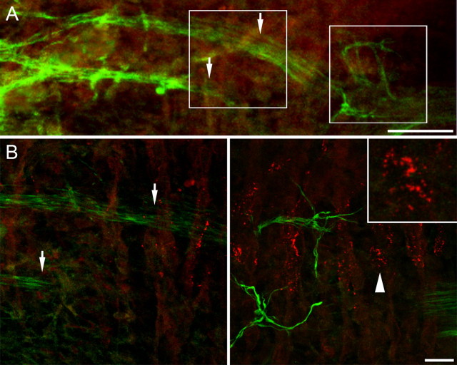
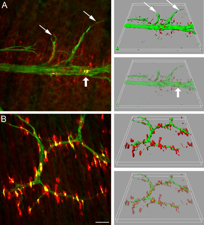
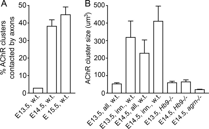
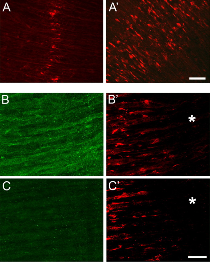
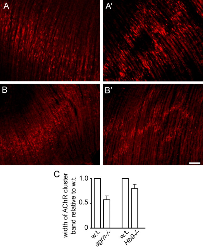
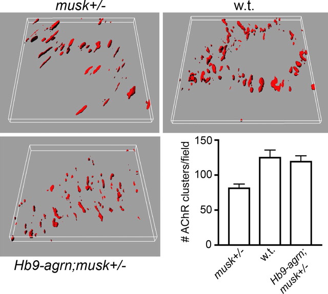
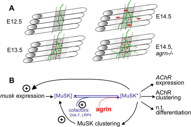
Neuromuscular junctions (NMJs) normally form in the central region of developing muscle. In this process, agrin released from motor neurons has been considered to initiate the formation of synaptic acetylcholine receptor (AChR) clusters (neurocentric model). However, in muscle developing in the absence of nerves and thus of agrin, AChR clusters still form in the muscle center. This raises the possibility that the region of NMJ formation is determined by muscle-derived cues that spatially restrict the nerve to form synapses from aneural AChR clusters, e.g., by patterned expression of the agrin receptor MuSK (muscle-specific kinase) (myocentric model). Here we examine at initial stages of synaptogenesis whether the responsiveness of myotubes to agrin is spatially restricted, whether the regions of NMJ formation in wild-type muscle and of aneural AChR cluster formation in agrin-deficient animals correlate, and whether AChR cluster growth depends on the presence of agrin. We show that primary myotubes form AChR clusters in response to exogenous agrin in their central region only, a pattern that can spatially restrict NMJ formation. However, the nerve also makes synapses in regions in which aneural AChR clusters do not form, and agrin promotes synaptic cluster growth from the first stages of neuromuscular contact formation. These data indicate that aneural AChR clusters per se are not required for NMJ formation. A model is proposed that explains either the neurocentric or the myocentric mode of NMJ formation depending on a balance between the levels of MuSK expression and the availability of nerve-released agrin.


Figures

Figure 1.
Figure 1.
Wild-type mouse diaphragm at E12.5. Maximum intensity projection of stacks of images taken at 2 μm (A) and 0.1 μm (B) intervals. Speckles of AChRs are present on the myotubes near but not distant from the area of nerve entry. They run in a band centered beneath the nerve, but none are in contact with it. Left and right frames in A denote the areas enlarged in left and right bottom B, respectively. Arrows mark nerve trunks in A and B, left. Close-up inset in B, right, shows microclusters grouped but not fused to a single AChR cluster (arrowhead). Scale bars: A, 70 μm; B and inset, 20 and 7 μm, respectively.
Figure 2.
Figure 2.
Wild-type mouse diaphragms at E13.5 (A) and E14.5 (B). In three consecutive frames of 240 × 240 μm in the vicinity of the nerve entry, stacks of images were taken at 0.5 μm intervals, and maximum intensity projections (left) and three-dimensional renderings (right, in full and semitransparent coloring) were made to resolve neuromuscular contacts (yellow). At E13.5 (A), microclusters increased in number and many are enlarged compared with E12.5. Only a small minority of clusters in the central AChR cluster band are contacted by nerve (large arrow). Note that some branches grow laterally beyond the AChR cluster band (small arrows). At E14.5 (B), the band of AChR patches is significantly wider and AChR patches are significantly larger than at E13.5. Numerous clusters are contacted by axons (yellow). Scale bar, 30 μm.
Figure 3.
Figure 3.
Numbers of AChR clusters contacted by nerves (in percentage of all clusters; A) and sizes of AChR clusters between E13.5 and E15.5 (B). Data from image stacks as illustrated in Figure 2. Note that the number of AChR clusters contacted by axons and their size increase strongly during this period. In contrast, no contacts are made when nerves do not express agrin, nor do clusters grow in the absence of nerves (Hb9−/−) or agrin (agrn−/−). Data on agrin mutants are from littermates. Each column gives mean ± SE from 11–16 stacks analyzed, taken from three to four animals, except Hb9−/− (3–6 stacks, 2 animals), each stack containing 51–556 AChR clusters.
Figure 4.
Figure 4.
The responsiveness of myotubes in E14.5 diaphragms to agrin is restricted to their central regions. A, A′, E14.5 diaphragms cultured for 18 h in the absence (A) and presence (A′) of 10 nm chicken mini-agrin cN257C21B8. Note that, despite the presence of agrin, clusters are confined to the central region of the myotubes. Scale bar, 40 μm. B, B′, Diaphragms of MCK–cmagB8 mice expressing mini-agrin from myotubes in vivo at E13.5. B, After staining for agrin immunoreactivity; B′, same region stained for AChR clusters. Note absence of AChR clusters in lateral regions (*) of the diaphragm despite uniform agrin transgene expression along entire myotubes. C, C′, Same as in B but from w.t. littermate. Scale bar, 25 μm.
Figure 5.
Figure 5.
The width of the central band of AChR clusters in diaphragms at E14.5 is narrower in agrn−/− (A) than in control (A′) littermates. A difference is also found between Hb9−/− mutants (B) and control (B′) littermates. C, Widths of AChR cluster bands (means ± SE) in agrn−/− and Hb9−/− mutants relative to control littermates. Data are based on five agrn−/− mutants (2 litters; p < 0.05) and four Hb9−/− mutants (3 litters; p < 0.1). For method, see supplemental Figure 2 (available at www.jneurosci.org as supplemental material). Scale bar, 70 μm.
Figure 6.
Figure 6.
Reduction in AChR cluster number in musk+/− mutants is compensated by overexpression of an agrin transgene in motor neurons. All panels are based on data from a single litter. n = 2 animals each for all genotypes, 11–12 stacks of optical sections taken at 40× in 0.5 μm steps from both hemidiaphragms of each animal. Differences in cluster numbers between musk +/− versus Hb9–agrn;musk+/− and w.t., respectively, are significant (p < 0.01).
Figure 7.
Figure 7.
A, Steps of NMJ formation in diaphragms between E12.5 and E14.5 and effects of agrn ablation (bottom right). Dark gray area marked by dotted lines denotes the region in which aneural AChR clusters form. Adjacent light gray region is responsive to agrin but does not form aneural AChR clusters, perhaps attributable to lower expression levels of MuSK and/or its accessory proteins, e.g., Dok-7 or LRP4. B, Model for early stages of neuromuscular synapse formation. The formation of AChR clusters depends on the local concentration of activated MuSK (MuSK*). In muscle regions in which MuSK is low, agrin from nerves is needed for MuSK activation, resulting in synapse formation consistent with the neurocentric model. Conversely, in regions expressing high levels of MuSK (and of cofactors such as Dok-7 and LRP4), MuSK is autoactivated (MuSK*) and forms numerous aneural AChR clusters. This results in a high probability of motor neurons encountering a preformed AChR cluster, thus favoring innervation of preformed aneural AChR clusters according to the myocentric model. Also shown are feedback loops by which MuSK* stimulates its own aggregation and expression (Jones et al., 1999; Moore et al., 2001).

Similar articles

Cited by

References

    1. Anderson MJ, Cohen MW. Nerve-induced and spontaneous redistribution of acetylcholine receptors on cultured muscle cells. J Physiol (Lond) 1977;268:757–773. - PMC - PubMed
    1. Arber S, Han B, Mendelsohn M, Smith M, Jessell TM, Sockanathan S. Requirement for the homeobox gene Hb9 in the consolidation of motor neuron identity. Neuron. 1999;23:659–674. - PubMed
    1. Ashby PR, Wilson SJ, Harris AJ. Formation of primary and secondary myotubes in aneural muscles in the mouse mutant peroneal muscular atrophy. Dev Biol. 1993a;156:519–528. - PubMed
    1. Ashby PR, Pinçon-Raymond M, Harris AJ. Regulation of myogenesis in paralyzed muscles in the mouse mutants peroneal muscular atrophy and muscular dysgenesis. Dev Biol. 1993b;156:529–536. - PubMed
    1. Babiuk RP, Zhang W, Clugston R, Allan DW, Greer JJ. Embryological origins and development of the rat diaphragm. J Comp Neurol. 2003;455:477–487. - PubMed

Publication types

MeSH terms