. 2008 Mar 26;28(13):3384-91.
doi: 10.1523/JNEUROSCI.0185-08.2008.

The chaperone activity of heat shock protein 90 is critical for maintaining the stability of leucine-rich repeat kinase 2

Affiliations

The chaperone activity of heat shock protein 90 is critical for maintaining the stability of leucine-rich repeat kinase 2

Lizhen Wang et al. J Neurosci. .

Abstract

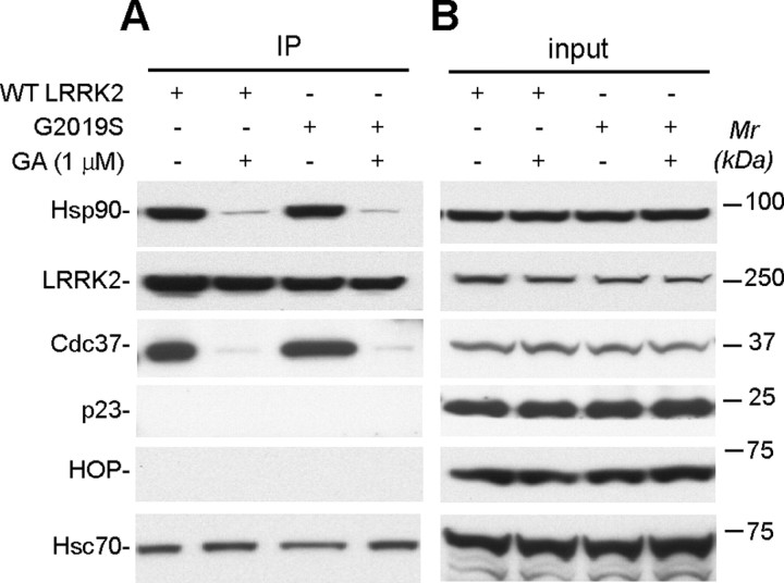
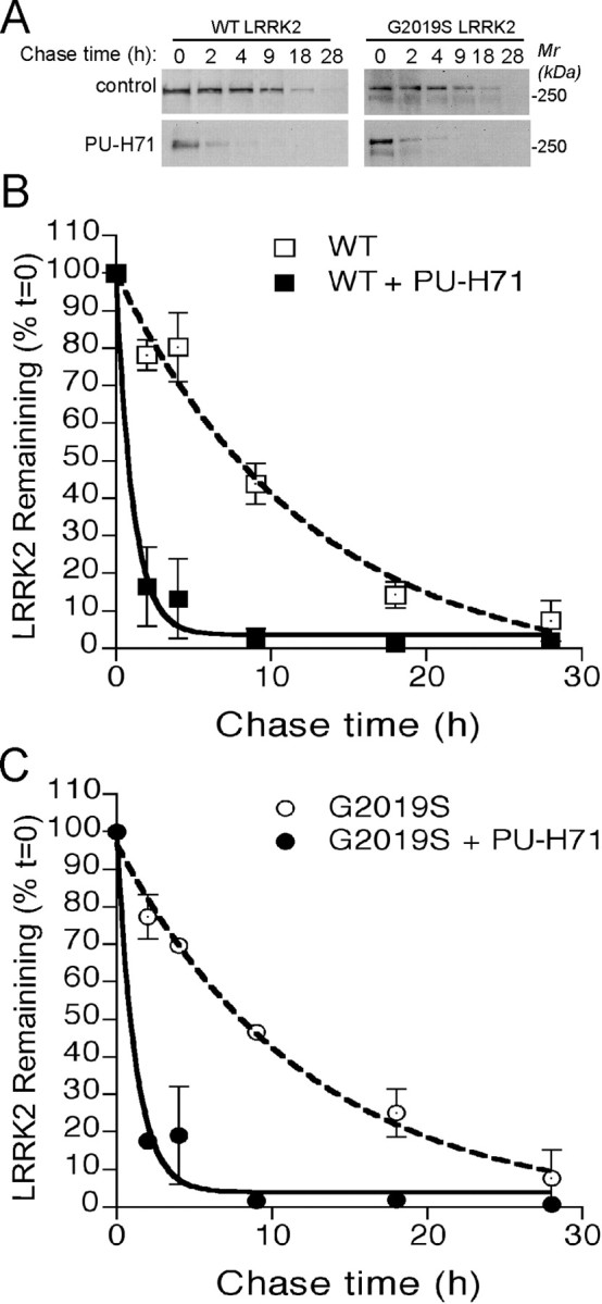
Parkinson's disease (PD), a progressive neurodegenerative disease characterized by bradykinesia, rigidity, and resting tremor, is the most common neurodegenerative movement disorder. Although the majority of PD cases are sporadic, some are inherited, including those caused by leucine-rich repeat kinase 2 (LRRK2) mutations. The substitution of serine for glycine at position 2019 (G2019S) in the kinase domain of LRRK2 represents the most prevalent genetic mutation in both familial and apparently sporadic cases of PD. Because mutations in LRRK2 are likely associated with a toxic gain of function, destabilization of LRRK2 may be a novel way to limit its detrimental effects. Here we show that LRRK2 forms a complex with heat shock protein 90 (Hsp90) in vivo and that inhibition of Hsp90 disrupts the association of Hsp90 with LRRK2 and leads to proteasomal degradation of LRRK2. Hsp90 inhibitors may therefore limit the mutant LRRK2-elicited toxicity to neurons. As a proof of principle, we show that Hsp90 inhibitors rescue the axon growth retardation caused by overexpression of the LRRK2 G2019S mutation in neurons. Therefore, inhibition of LRRK2 kinase activity can be achieved by blocking Hsp90-mediated chaperone activity and Hsp90 inhibitors may serve as potential anti-PD drugs.


Figures

Figure 1.
Figure 1.
LRRK2 complexes with Hsp90 in LRRK2 G2019S mutant transgenic mouse brains. A, Silver staining of purified LRRK2-containing protein complexes from LRRK2 G2019S transgenic (G2019S) mouse brains. Protein bands seen in LRRK2 G2019S but not in ntg mouse brain samples were subjected to mass spectrometry analysis. LRRK2 and Hsp90 chaperone proteins were selectively identified in LRRK2 immunoprecipitates. B–D, LRRK2 was immunoprecipitated with HA matrix from G2019S and ntg mouse brains, and the immunoprecipitates (IP) were blotted with antibodies against Hsp90, Cdc37, and Hsp70. Hsp90 and Cdc37, but not Hsp70, were coimmunoprecipitated with LRRK2 from mouse brain extracts. Mr, Molecular weight; WB, Western blots.
Figure 2.
Figure 2.
Hsp90 inhibitor disrupts the LRRK2/Hsp90 complex. A, HEK-293 cells transfected with either WT or G2019S mutant LRRK2 were treated with Hsp90 inhibitor GA (+) or vehicle (−) for 1 h, lysed, immunoprecipitated with HA matrix, and then subjected to Western blot analysis with antibodies against Hsp90, LRRK2, Cdc37, p23, HOP, and Hsc70. B, The expression of Hsp90, LRRK2, Cdc37, p23, HOP, and Hsc70 in cell extracts was revealed by Western blot analysis with antibodies against Hsp90, Cdc37, p23, HOP, and Hsc70. LRRK2 was detected by the anti-HA antibody. IP, Immunoprecipitates; Mr, molecular weight.
Figure 3.
Figure 3.
Hsp90 inhibitors suppress the steady level of LRRK2 but not α-synuclein expression in HEK-293 cells. A, B, Western blot analysis of LRRK2 expression in HEK-293 cells transfected with G2019S mutant (G2019S) (A) and WT LRRK2 after treatment with the indicated doses of Hsp90 inhibitor PU-H71 for 24 h (B). C, Dose–response curve of overexpressed wild-type (filled triangles) and G2019S mutant (open triangles) LRRK2 protein treated with PU-H71. D, Western blot analysis of α-synuclein A53T expression in transfected HEK-293 cells treated with vehicle (DMSO) or 500 nm GA for 24 h. Mr, Molecular weight.
Figure 4.
Figure 4.
Hsp90 inhibitors suppress the steady level of mutant and endogenous LRRK2 in neurons. A, B, Western blot analysis of LRRK2 expression in primary cultured cortical neurons derived from LRRK2 G2019S transgenic (G2019S) and littermate nontransgenic pups (endogenous) after treatment of indicated doses of Hsp90 inhibitor GA for 24 h (A) or 10 nm GA for indicated time points (B). C, D, Dose–response curves of endogenous (filled triangles) and G2019S mutant (open triangles) LRRK2 protein treated with GA (C) or PU-H71 (D). LRRK2 was detected by a LRRK2 polyclonal antibody, JH5514 (Biskup et al., 2006). Mr, Molecular weight.
Figure 5.
Figure 5.
Hsp90 regulates the stability of LRRK2. A, WT and G2019S mutant LRRK2 proteins were pulse labeled with biotinylated Halo ligand and chased at indicated time intervals in the presence or absence (control) of 1 μm PU-H71. B, C, Time courses of the decay of biotinylated WT (B) and G2019S mutant (C) LRRK2 proteins in the presence or absence (control) of Hsp90 inhibitor PU-H71. Mr, Molecular weight.
Figure 6.
Figure 6.
Hsp90 inhibitor-induced suppression of LRRK2 expression is mediated by the proteasome-dependent protein degradation pathway. A, B, Western blot analyses of LRRK2 protein in primary cultured cortical neurons derived from LRRK2 G2019S mutant (A) and ntg (B) pups after treatment with Hsp90 inhibitor GA in the presence or absence of the proteasome inhibitor MG132 (MG) or the lysosome inhibitor chloroquine (Ch). LRRK2 was detected by an LRRK2 polyclonal antibody, JH5514. Mr, Molecular weight.
Figure 7.
Figure 7.
PU-H71 rescues the axonal growth deficit of neurons derived from LRRK2 G2019S mutant mice. A–D, Representative images of hippocampal neurons derived from nontransgenic (A) and LRRK2 G2019S transgenic (C, D) pups treated with vehicle (Veh), doxycycline (Dox), or PU-H71 for 2 d and visualized by immunostaining of βIII-tubulin antibody. Scale bar, 100 μm. E, F, Bar graphs of axon length of LRRK2 G2019S (E) and nontransgenic (F) neurons after 2 d in culture. Data are presented as means ± SEM. *p < 0.0001.

Similar articles

Cited by

References

    1. Auluck PK, Bonini NM. Pharmacological prevention of Parkinson disease in Drosophila. Nat Med. 2002;8:1185–1186. - PubMed
    1. Auluck PK, Meulener MC, Bonini NM. Mechanisms of suppression of α-synuclein neurotoxicity by geldanamycin in Drosophila. J Biol Chem. 2005;280:2873–2878. - PubMed
    1. Biskup S, Moore DJ, Celsi F, Higashi S, West AB, Andrabi SA, Kurkinen K, Yu SW, Savitt JM, Waldvogel HJ, Faull RL, Emson PC, Torp R, Ottersen OP, Dawson TM, Dawson VL. Localization of LRRK2 to membranous and vesicular structures in mammalian brain. Ann Neurol. 2006;60:557–569. - PubMed
    1. Bonifati V. Parkinson's disease: the LRRK2–G2019S mutation: opening a novel era in Parkinson's disease genetics. Eur J Hum Genet. 2006;14:1061–1062. - PubMed
    1. Cai H, Lin X, Xie C, Laird FM, Lai C, Wen H, Chiang HC, Shim H, Farah MH, Hoke A, Price DL, Wong PC. Loss of ALS2 function is insufficient to trigger motor neuron degeneration in knock-out mice but predisposes neurons to oxidative stress. J Neurosci. 2005;25:7567–7574. - PMC - PubMed

Publication types

MeSH terms