. 2008 Mar 26;28(13):3404-14.
doi: 10.1523/JNEUROSCI.0157-08.2008.

A molecular platform in neurons regulates inflammation after spinal cord injury

Affiliations

A molecular platform in neurons regulates inflammation after spinal cord injury

Juan Pablo de Rivero Vaccari et al. J Neurosci. .

Abstract

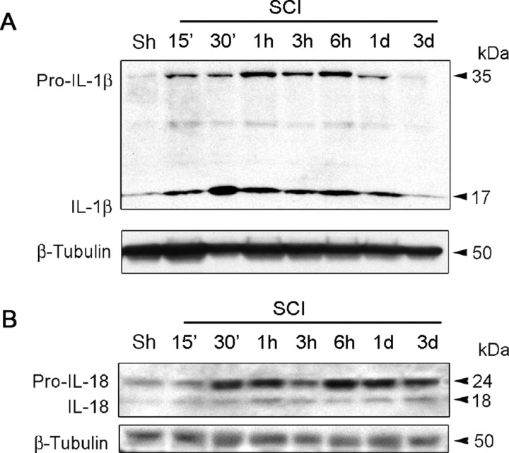
Vigorous immune responses are induced in the immune privileged CNS by injury and disease, but the molecular mechanisms regulating innate immunity in the CNS are poorly defined. The inflammatory response initiated by spinal cord injury (SCI) involves activation of interleukin-1beta (IL-1beta) that contributes to secondary cell death. In the peripheral immune response, the inflammasome activates caspase-1 to process proinflammatory cytokines, but the regulation of trauma-induced inflammation in the CNS is not clearly understood. Here we show that a molecular platform [NALP1 (NAcht leucine-rich-repeat protein 1) inflammasome] consisting of caspase-1, caspase-11, ASC (apoptosis-associated speck-like protein containing a caspase-activating recruitment domain), and NALP1 is expressed in neurons of the normal rat spinal cord and forms a protein assembly with the X-linked inhibitor of apoptosis protein (XIAP). Moderate cervical contusive SCI induced processing of IL-1beta, IL-18, activation of caspase-1, cleavage of XIAP, and promoted assembly of the multiprotein complex. Anti-ASC neutralizing antibodies administered to injured rats entered spinal cord neurons via a mechanism that was sensitive to carbenoxolone. Therapeutic neutralization of ASC reduced caspase-1 activation, XIAP cleavage, and interleukin processing, resulting in significant tissue sparing and functional improvement. Thus, rat spinal cord neurons contain a caspase-1, pro-ILbeta, and pro-IL-18 activating complex different from the human NALP1 inflammasome that constitutes an important arm of the innate CNS inflammatory response after SCI.


Figures

Figure 1.
Figure 1.
SCI induces processing of IL-1β and IL-18 early after injury. Representative immunoblot analysis of spinal cord lysates of sham animals (Sh) and traumatized rat cords at 15 min, 30 min, 1 h, 3 h, 6 h, 1 d, and 3 d after injury. A, B, Spinal cord lysates were immunoblotted with antibodies against IL-1β (A) and IL-18 (B). β-Tubulin was used as internal standard and control for protein loading.
Figure 2.
Figure 2.
SCI induces activation and processing of caspase-1 and increases levels of ASC and caspase-11 but not NALP1. A–D, Representative immunoblot analysis of caspase-1 (A), caspase-11 (B), ASC (C), and NALP1 (D) in spinal cord lysates of sham (Sh) and traumatized rat cords at indicated times after injury. C shows that anti-ASC reacts specifically with ASC (26 kDa) and nonspecifically with proteins of ∼49 and 55 kDa (arrows, left). β-Tubulin was used as internal standard and control for protein loading. Data are presented as mean ± SEM. *p < 0.05, #p < 0.10 compared with sham. n = 5 per group.
Figure 3.
Figure 3.
SCI induces association of NALP1 inflammasome proteins, processing of caspase-1, and cleavage of XIAP. Coimmunoprecipitation with ASC of sham lysates (Sh) and lysates obtained at 30 min, 6 h, and 3 d after SCI. ASC immunoprecipitates were blotted for ASC, caspase-1, caspase-11, NALP1, XIAP, and caspase-3 (control). In sham animals, anti-ASC immunoprecipitated (IP) NALP1, caspase-1, caspase-11, and the 53 kDa XIAP, thus indicating association of these proteins in a multiprotein complex. SCI induced increased association of NALP1 inflammasome proteins, processing of caspase-1 and caspase-11, and cleavage of XIAP into fragments. Preimmune serum did not immunoprecipitate inflammasome proteins and was used as control.
Figure 4.
Figure 4.
NALP1 inflammasome proteins are present in spinal cord motor neurons, and SCI induces alterations in protein expression patterns. Confocal images show motor neurons in the ventral horn of sham and injured spinal cords at 6 h after trauma. Sections were stained for caspase-1, caspase-11, ASC, and NALP1 (red) and the neuronal marker MAP2 (green). In sham animals, caspase-1 immunoreactivity was seen in the nucleus (arrow). By 6 h after injury, increased caspase-1 staining was present in neuronal nuclei, and patchy staining was present in the cell cytoplasm (arrow) and processes near the plasma membrane. Caspase-11 immunoreactivity showed diffuse punctate staining confined to the neuronal soma and processes (arrow). Increased caspase-11 staining was present by 6 h after trauma in the neuronal soma in a patchy distribution (arrow). Intense ASC and NALP1 staining was detected in the soma of spinal cord neurons and exhibited a patchy distribution pattern in the cytoplasm (arrow). Both inflammasome proteins showed increased expression as evidenced by intense patchy staining located near or associated with the plasma membrane (arrows) by 6 h after trauma. Scale bars, 20 μm.
Figure 5.
Figure 5.
A, NALP1 inflammasome proteins are expressed in spinal cord neurons in culture. Spinal cord neurons (∼95% pure) were grown in culture for 14 d, harvested, and lysed. A segment (C5–C7) of adult rat spinal cord was excised and homogenized as outlined in Materials and Methods. Samples were immunoblotted for NALP1, ASC, caspase-1, caspase-11, and XIAP. Neurons in culture (culture); spinal cord tissue (cord). B, Activation of caspase-1 is induced in spinal cord neurons by treatment with the K+ ionophore valinomycin. Spinal cord neuronal cultures were grown for 14 d and treated for 4 h with 1 μm valinomycin or were left untreated (control). The cells were lysed and analyzed by immunoblot for caspase-1.
Figure 6.
Figure 6.
A, Confocal images of spinal cord sections demonstrating that anti-ASC antibody is taken up by spinal cord neurons. Anti-ASC was conjugated to FITC using the EZ-Label FITC Protein Labeling kit (Pierce) according to the instructions of the manufacturer. Rats were subjected to moderate cervical SCI. At 20 min after SCI, 50 μg of anti-ASC–FITC was injected intraperitoneally and intravenously. Control injured rats received FITC alone. At 6 h after SCI, anti-ASC–FITC labeled cervical motor neurons in the ventral horn, whereas these cells were not labeled by administration of FITC alone. The right panel (ASC) shows spinal cord sections immunostained with anti-ASC followed by appropriate secondary antibody for comparison. Scale bars, 20 μm. B, Incorporation of anti-ASC into spinal cord neurons is sensitive to carbenoxolone. Spinal cord neurons were grown for 7 d and treated with anti-ASC–FITC (ASC), anti-actin–FITC (actin), and IgG–FITC (IgG). Other cultures were pretreated for 10 min with 100 μm carbenoxolone and anti-ASC (CBX) or 30 μm cytochalasin D (CytochD) and then incubated with anti-ASC for 1 h. FITC-conjugated anti-ASC was taken up by spinal cord neurons, whereas FITC-conjugated actin and IgG were excluded from these cells. Incorporation of anti-ASC–FITC was blocked by carbenoxolone, whereas cytochalasin D had little effect. The bottom row shows DAPI-labeled nuclei corresponding to cells in top row.
Figure 7.
Figure 7.
ASC neutralization decreases SCI-induced activation and processing of caspase-1, IL-1β, IL-18, and XIAP cleavage. Representative immunoblots of injured spinal cords from animals subjected to SCI and treated intraperitoneally and intravenously with antibodies to ASC (A), IgG controls (IgG), or were left untreated (N) at 20 min after injury. Animals were killed 24 h after treatment. Treatment resulted in inhibition of inflammasome activation as detected by a decrease in the processing of procaspase-1, cleavage of XIAP, and a reduction in the levels of cleaved IL-1β and IL-18.
Figure 8.
Figure 8.
ASC neutralization improves histopathological outcome and decreases spinal cord lesion volume. A, Representative cross sections of spinal cords of antibody-treated (Anti-ASC) and nontreated (No Antibody) animals at 3 d after SCI. Hematoxylin–eosin and luxol fast blue stained sections represent the injury epicenter and sites 4.2 mm rostral and caudal to the epicenter. Administration of anti-ASC significantly reduced the lesion volume at 3 d after injury. Areas of degeneration evaluated for volumetric analysis of lesion volume were determined by diminished white matter degeneration and preservation of motor neuron morphology. Significance was determined by comparing average lesion volume of antibody-treated animals to control groups using Student's t test (n = 5 per group). There were no differences between nontreated animals (no antibody) and IgG-treated controls. B, Spinal cords from animals treated with anti-ASC demonstrated smaller areas of shrunken neurons in gray matter (arrowheads) and reduced white matter degeneration (arrows). Scale bar, 50 μm.
Figure 9.
Figure 9.
ASC neutralization improves functional outcomes after SCI. A–E, After SCI, a person blinded to the treatment protocols assessed behavior with the following tests: gripping force (A), sticker removal (B), footprint analysis/base of support (C), stride length (D), and foot rotation (E). Data are presented as the mean ± SD. *p < 0.05 compared with sham.

References

    1. Agostini L, Martinon F, Burns K, McDermott MF, Hawkins PN, Tschopp J. NALP3 forms an IL-1beta-processing inflammasome with increased activity in Muckle-Wells autoinflammatory disorder. Immunity. 2004;20:319–325. - PubMed
    1. Bareyre FM, Schwab ME. Inflammation, degeneration and regeneration in the injured spinal cord: insights from DNA microarrays. Trends Neurosci. 2003;26:555–563. - PubMed
    1. Bartholdi D, Schwab ME. Expression of pro-inflammatory cytokine and chemokine mRNA upon experimental spinal cord injury in mouse: an in situ hybridization study. Eur J Neurosci. 1997;9:1422–1438. - PubMed
    1. Benveniste EN. Inflammatory cytokines within the central nervous system: sources, function, and mechanism of action. Am J Physiol. 1992;263:C1–C16. - PubMed
    1. Bethea JR. Spinal cord injury-induced inflammation: a dual-edged sword. Prog Brain Res. 2000;128:33–42. - PubMed

Publication types

MeSH terms