. 2008 Aug;22(8):2821-31.
doi: 10.1096/fj.07-104224. Epub 2008 Mar 26.

G alpha12 is targeted to the mitochondria and affects mitochondrial morphology and motility

Affiliations

G alpha12 is targeted to the mitochondria and affects mitochondrial morphology and motility

Alexandra V Andreeva et al. FASEB J. 2008 Aug.

Abstract

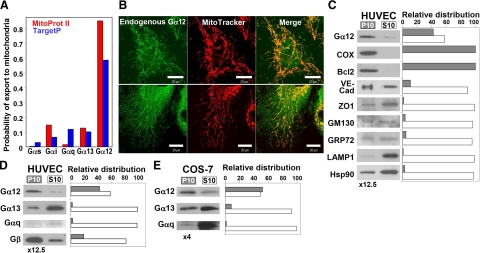
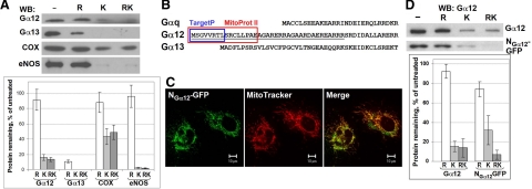
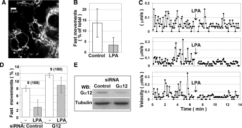
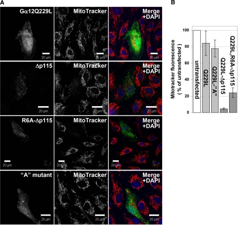
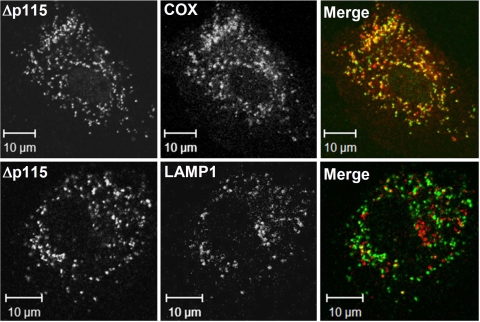
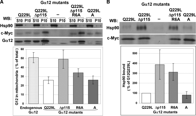
G alpha12 constitutes, along with G alpha13, one of the four families of alpha subunits of heterotrimeric G proteins. We found that the N terminus of G alpha12, but not those of other G alpha subunits, contains a predicted mitochondrial targeting sequence. Using confocal microscopy and cell fractionation, we demonstrated that up to 40% of endogenous G alpha12 in human umbilical vein endothelial cells colocalize with mitochondrial markers. N-terminal sequence of G alpha12 fused to GFP efficiently targeted the fusion protein to mitochondria. G alpha12 with mutated mitochondrial targeting sequence was still located in mitochondria, suggesting the existence of additional mechanisms for mitochondrial localization. Lysophosphatidic acid, one of the known stimuli transduced by G alpha12/13, inhibited mitochondrial motility, while depletion of endogenous G alpha12 increased mitochondrial motility. G alpha12Q229L variants uncoupled from RhoGEFs (but not fully functional activated G alpha12Q229L) induced transformation of the mitochondrial network into punctate mitochondria and resulted in a loss of mitochondrial membrane potential. All examined G alpha12Q229L variants reduced phosphorylation of Bcl-2 at Ser-70, while only mutants unable to bind RhoGEFs also decreased cellular levels of Bcl-2. These G alpha12 mutants were also more efficient Hsp90 interactors. These findings are the first demonstration of a heterotrimeric G protein alpha subunit specifically targeted to mitochondria and involved in the control of mitochondrial morphology and dynamics.


Figures

Figure 1.
Figure 1.
Endogenous Gα12 is localized to mitochondria in HUVECs and COS-7 cells. A) Prediction of mitochondrial targeting of different Gα subunits using Mitoprot II (red) and TargetP (blue). UniProt accession numbers: Gαs, P63092; Gαi1, P63096; Gαq, P50148; Gα12, Q03113; Gα13, Q14344. B) Colocalization of endogenous Gα12 with MitoTracker in HUVECs. Living cells were incubated with MitoTracker, fixed, and immunostained with Gα12-specific antibodies (top and bottom panels: lots H052 and I150, respectively). Scale bars = 20 μm. C) Distribution of Gα12 and organelle markers in the mitochondrial fraction (P10) and the fraction containing other organelles except nuclei (S10). P10 was resuspended in 1/12.5 of initial volume, and equal volumes of P10 and S10 were loaded on the gel. The following marker proteins were examined: COX and Bcl-2 (mitochondria), VE-cadherin, and zonula occludens-1 (ZO1) (plasma membrane/microsomal fraction), GM130 (Golgi apparatus), GRP72 (endoplasmic reticulum), LAMP1 (lysosomes), as well as Hsp90 as a representative of proteins mainly present in the cytoplasm. D, E) Distribution of subunits of different heterotrimeric G proteins in HUVECs and in COS-7 cells, respectively. In E, P10 was resuspended in 1/4 of initial volume. Experiments shown were repeated twice with similar results.
Figure 2.
Figure 2.
N terminus of Gα12 targets proteins to the mitochondrial surface. A) Mitochondrial fraction of HUVECs was subjected to hypotonic rupture (R) and/or treated with Proteinase K (K) as indicated. The effect of these treatments on Gα12, Gα13, COX, and eNOS was assessed by Western blotting with corresponding antibodies. Data were normalized to the protein content in untreated aliquots. Data shown are the means of two replicates; error bars show values obtained in each replicate. B) Sequences in Gα12 predicted by Mitoprot II (red) and TargetP (blue) to function as signal peptide for mitochondrial targeting. UniProt accession numbers: Gαq, P21279; Gα12, P27600; Gα13, P27601. C) Mitochondrial targeting of NGα12-GFP (GFP fused to the N terminus of Gα12, underlined in B). Twenty-four hours after transfection, living HUVECs were incubated with MitoTracker and examined using confocal microscopy. D) N terminus of Gα12 does not mediate GFP translocation into mitochondrial matrix. Mitochondrial fraction obtained from COS-7 cells (either nontransfected or transfected with NGα12-GFP) was subjected to Proteinase K protection assay as in A. Endogenous Gα12 and NGα12-GFP were detected by Western blotting with Gα12-specific antibody. Data are means of 3 replicates; error bars indicate sd.
Figure 3.
Figure 3.
LPA reduces (A–C) and Gα12 depletion (D–E) increases mitochondrial motility in HUVECs. A–C) HUVECs were starved for 24 h and incubated with MitoTracker to visualize mitochondria. A) The first frame of the recorded image sequence. Movement of 27 individual mitochondria was tracked under control conditions. Those that showed fast movement (exceeding the threshold of 0.1 μm/s) were also tracked after LPA addition (7 mitochondria marked with circles). Scale bar = 5 μm. B) Percentage of fast movements during entire recording before (control) and after (LPA) addition of LPA. Data are means ± sd; P = 0.011, paired Student’s t test. C) Representative plots of velocity vs. time for 3 individual mitochondria. Arrows indicate LPA addition. D–E) HUVECs were transfected with Gα12 siRNA duplexes or control siRNA. Mitochondrial movement was tracked 24 h after transfection for 6 min before and for additional 6 min after LPA addition. Mitochondrial movement was quantified as the percentage of mitochondria moving faster than 0.1 μm/s at any time point during recording. D) Quantification data shown are the means of two replicates (two separate slides), with error bars showing values obtained in each replicate. Data indicate total number of cells and number of individual mitochondria (in parentheses). The extent of Gα12 depletion was assessed by Western blotting using α-tubulin as a loading control (E).
Figure 4.
Figure 4.
Gα12 mutants unable to activate Rho lead to mitochondrial fragmentation and to an increased mitochondrial permeability. HUVECs were transfected with the Myc-tagged Gα12Q226L, Gα12Q229L-Δp115RhoGEF, Gα12Q229L,R6A-Δp115RhoGEF, and Gα12Q229L A (with residues 4–8 replaced with a NAAIR sequence; ref. ; constructs as indicated). A) 24 h after transfection, cells were incubated with MitoTracker, fixed, and immunostained with Myc-specific antibody. Scale bars = 20 μM. B) MitoTracker-labeled mitochondria in transfected and surrounding untransfected cells were quantified using ImageJ. Fifteen transfected cells for each Gα12 mutant were analyzed. Error bars indicate sd.
Figure 5.
Figure 5.
Punctate structures induced by Rho-uncoupled Gα12Q229L are positive for a mitochondrial (COX) rather than late endosomal/lysosomal (LAMP1) marker. Experiment was performed as in Fig. 4, and cells were stained with Myc-specific antibody (Δp115) and polyclonal antibodies against COX and LAMP1 as indicated.
Figure 6.
Figure 6.
Mitochondrial targeting of Gα12Q229L mutants and their interaction with Hsp90. A) COS-7 cells were transfected with either pcDNA3.1-Myc-His6, or Gα12Q229L-Myc constructs as indicated. Cells were harvested 48 h after transfection and fractionated into mitochondrial pellet (P10) and supernatant (S10). Fractions were probed by Western blotting for the presence of Hsp90 and Gα12 (the latter detected using either c-Myc or Gα12 antibody; note that mutations in the R6A and A constructs are within the epitope of Gα12 antibody and are likely to affect its ability to recognize respective proteins). B) Mitochondrial fractions were solubilized in 0.5% Nonidet P-40; their amounts were adjusted to contain similar amounts of Myc-tagged Gα12; and immunoprecipitation was performed using c-Myc antibody. The presence of Myc-tagged Gα12 and Hsp90 in immunoprecipitates was detected using respective antibodies. Bottom panel shows quantification of the Western blotting data for Hsp90 binding, normalized to c-Myc signal in respective samples. ECL quantification data are means of two independent experiments; error bars indicate values obtained in each experiment.
Figure 7.
Figure 7.
Effects of Gα12Q229L mutants on JNK and Bcl-2. COS-7 cells were transfected with either pcDNA3.1-Myc-His6 or Gα12Q229L-Myc constructs as indicated. A) The presence and phosphorylation levels of JNK (p46) and Bcl-2 (phosphorylated at Ser-70), as well as Myc-tagged Gα12 and COX (as a mitochondrial marker) were detected by Western blotting using respective antibodies (c, control). The diagrams show quantification of the ECL data shown in A: effects of Gα12Q229L-Myc mutants on phosphorylation state of JNK (p46) (B), or Bcl-2 (C), or on the cellular levels of Bcl-2 (D). Data are means of 3 replicates; error bars indicate sd. The experiment was repeated twice (3 replicates in each experiment) with similar results.

References

    1. DiMauro S, Schon E A. Mitochondrial respiratory-chain diseases. N Engl J Med. 2003;348:2656–2668. - PubMed
    1. Schultz B E, Chan S I. Structures and proton-pumping strategies of mitochondrial respiratory enzymes. Annu Rev Biophys Biomol Struct. 2001;30:23–65. - PubMed
    1. Orrenius S, Gogvadze A, Zhivotovsky B. Mitochondrial oxidative stress: implications for cell death. Annu Rev Pharmacol. 2007;47:143–183. - PubMed
    1. Hollenbeck P J, Saxton W M. The axonal transport of mitochondria. J Cell Sci. 2005;118:5411–5419. - PMC - PubMed
    1. McBride H M, Neuspiel M, Wasiak S. Mitochondria: more than just a powerhouse. Curr Biol. 2006;16:R551–560. - PubMed

Publication types

MeSH terms

Substances

LinkOut - more resources