. 2008 Mar;4(1):73-8.
doi: 10.1007/s11302-008-9095-1. Epub 2008 Feb 8.

The inflammatory effects of UDP-glucose in N9 microglia are not mediated by P2Y14 receptor activation

Affiliations

The inflammatory effects of UDP-glucose in N9 microglia are not mediated by P2Y14 receptor activation

Vielska M Brautigam et al. Purinergic Signal. 2008 Mar.

Abstract

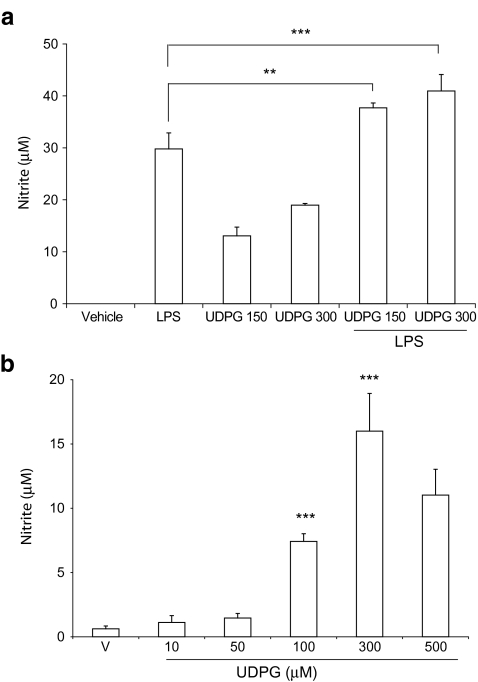
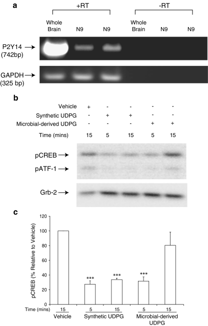
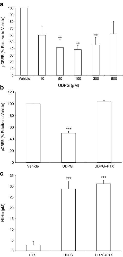
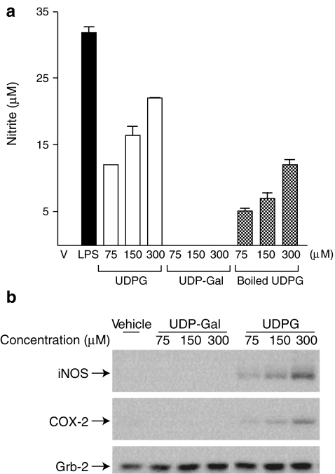
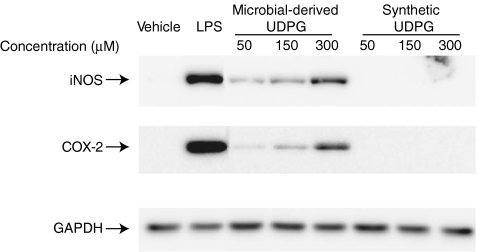
In this study we evaluated the functionality and inflammatory effects of P2Y14 receptors in murine N9 microglia. The selective P2Y14 receptor agonist UDP-glucose (UDPG) derived from microbial sources dose dependently stimulated expression of cyclooxygenase-2 and inducible nitric oxide synthase, and potentiated the effects of bacterial lipopolysaccharide on nitric oxide production. However, another selective P2Y14 receptor agonist, UDP-galactose, did not affect these endpoints either alone or in combination with lipopolysaccharide. Interestingly, synthetic UDPG also had no detectable pro-inflammatory effects, although P2Y14 receptors are both expressed and functional in N9 microglia. While synthetic UDPG decreased levels of phosphorylated cyclic AMP response element binding protein, an effect that was blocked by pertussis toxin, the pro-inflammatory effects of microbial-derived UDPG were insensitive to pertussis toxin. These data suggest that the pro-inflammatory effects of microbial-derived UDPG are independent of P2Y14 receptors and imply that microbial-derived contaminants in the UDPG preparation may be involved in the observed inflammatory effects.


Figures

Fig. 1
Fig. 1
UDPG potentiates LPS-stimulated NO release from N9 microglia. a Microglia were stimulated with vehicle (250 mM Hepes), LPS (1 μg/ml), UDPG (150 and 300 μM), or LPS and UDPG together for 18–22 h. Nitrite levels were determined using the Griess reagent. The graph is representative of at least five independent experiments performed in triplicate. b Microglia were treated with microbial-derived UDPG at the indicated concentrations for 18–22 h and nitrite levels assessed. **P < 0.01, ***P < 0.001
Fig. 2
Fig. 2
P2Y14 receptors are expressed and functional in N9 microglia. a Total RNA was isolated from two independently cultured N9 microglial thaws and from mouse whole brain as a positive control. The reverse transcription reaction was performed with (+) or without (−) reverse transcriptase (RT) followed by PCR amplification of P2Y14 mRNA (upper panel) or GAPDH mRNA (lower panel) as a control. b Microglia were stimulated in triplicate for 5 or 15 min with vehicle (250 mM Hepes), synthetic UDPG (300 μM), or microbial-derived UDPG (300 μM). Proteins from triplicate treatments were pooled and immunoblot analyses performed for phosphorylated CREB. Membranes were re-probed for the presence of Grb-2 as a loading control. The figure is representative of at least three independent experiments each performed in triplicate. c Quantification of phosphorylated CREB (pCREB) immunoreactivity obtained from the densitometric analyses of immunoblot studies (n ≥ 3) performed as shown in b. Normalized values (using Grb-2 immunoreactivity as a loading control) are expressed as percent CREB phosphorylation relative to vehicle treatment, and the data represent the mean ± SD of at least three separate experiments. **P < 0.01 vs. vehicle
Fig. 3
Fig. 3
Reductions in CREB phosphorylation stimulated by synthetic UDPG are sensitive to PTX, whereas microbial-derived UDPG-stimulated NO production is PTX insensitive. Quantification of phosphorylated CREB immunoreactivity obtained from the densitometric analyses of immunoblot studies (n ≥ 3). a Microglia were stimulated in triplicate with synthetic UDPG at the concentrations indicated for 15 min. b Microglia were pretreated with pertussis toxin (PTX; 100 ng/mL) for 18 h prior to stimulation with synthetic UDPG for 15 min. Proteins from triplicate treatments were pooled and immunoblot analyses performed for phosphorylated CREB. Membranes were re-probed for the presence of Grb-2 as a loading control. Normalized numerical values (using Grb-2 immunoreactivity as a loading control) are expressed as percent CREB phosphorylation relative to vehicle treatment, and the data represent the mean ± SD of at least three separate experiments. **P < 0.01 and ***P < 0.001 vs. vehicle. c Microglia were pretreated in triplicate with vehicle (PBS) or PTX (100 ng/mL) for 18 h prior to stimulation with either vehicle (250 mM Hepes) or UDPG (300 μM) for 18–22 h. Nitrite levels were determined using the Griess reagent. The graph represents cumulative quantification of at least three independent experiments, wherein treatments were performed in triplicate. ***P < 0.001 vs. PTX alone
Fig. 4
Fig. 4
The P2Y14-receptor ligand UDP-glucose, but not UDP-galactose, is pro-inflammatory in N9 microglia. Microglia were stimulated in triplicate with vehicle (V; 250 mM Hepes), UDPG (75, 150, or 300 μM), UDP-gal (75, 150, or 300 μM), or boiled UDPG (75, 150, or 300 μM) for 18–22 h. a Nitrite levels in the medium were determined using the Griess reagent. b Proteins were harvested, pooled and analyzed by immunoblot analyses for iNOS (upper panel) and COX-2 (middle panel). Grb-2 was used to control for protein loading (lower panel). The data shown are representative of at least three independent experiments
Fig. 5
Fig. 5
Microbial-derived UDPG stimulates iNOS and COX-2, whereas synthetic UDPG does not. Microglia were stimulated in triplicate for 18–22 h with either vehicle (250 mM Hepes), synthetic UDPG (50, 150, or 300 μM) or microbial-derived UDPG (50, 150, or 300 μM). Proteins from triplicate treatments were pooled and immunoblot analyses were performed for iNOS and COX-2 protein levels. Membranes were re-probed for the presence of GAPDH as a loading control. The figure is representative of at least three independent experiments performed in triplicate

References

    1. {'text': '', 'ref_index': 1, 'ids': [{'type': 'DOI', 'value': '10.1016/S0165-6147(02)00038-X', 'is_inner': False, 'url': 'https://doi.org/10.1016/s0165-6147(02)00038-x'}, {'type': 'PMC', 'value': 'PMC8653507', 'is_inner': False, 'url': 'https://pmc.ncbi.nlm.nih.gov/articles/PMC8653507/'}, {'type': 'PubMed', 'value': '12559763', 'is_inner': True, 'url': 'https://pubmed.ncbi.nlm.nih.gov/12559763/'}]}
    2. Abbracchio MP, Boeynaems J-M, Barnard EA, Boyer JL, Kennedy C, Miras-Portugal MT, King BF, Gachet C, Jacobson KA, Weisman GA, Burnstock G (2003) Characterization of the UDP-glucose receptor (re-named here the P2Y14 receptor) adds diversity to the P2Y receptor family. Trends Pharmacol Sci 24(2):52 - PMC - PubMed
    1. {'text': '', 'ref_index': 1, 'ids': [{'type': 'DOI', 'value': '10.1210/en.2004-0619', 'is_inner': False, 'url': 'https://doi.org/10.1210/en.2004-0619'}, {'type': 'PubMed', 'value': '15256495', 'is_inner': True, 'url': 'https://pubmed.ncbi.nlm.nih.gov/15256495/'}]}
    2. Baker AE, Brautigam VM, Watters JJ (2004) Estrogen modulates microglial inflammatory mediator production via interactions with estrogen receptor {beta}. Endocrinology 145(11):5021–5032 - PubMed
    1. {'text': '', 'ref_index': 1, 'ids': [{'type': 'DOI', 'value': '10.1046/j.1460-9568.2003.02663.x', 'is_inner': False, 'url': 'https://doi.org/10.1046/j.1460-9568.2003.02663.x'}, {'type': 'PubMed', 'value': '12814360', 'is_inner': True, 'url': 'https://pubmed.ncbi.nlm.nih.gov/12814360/'}]}
    2. Boucsein C, Zacharias R, Farber K, Pavlovic S, Hanisch UK, Kettenmann H (2003) Purinergic receptors on microglial cells: functional expression in acute brain slices and modulation of microglial activation in vitro. Eur J Neurosci 17(11):2267–2276 - PubMed
    1. {'text': '', 'ref_index': 1, 'ids': [{'type': 'DOI', 'value': '10.1016/j.jneuroim.2005.05.012', 'is_inner': False, 'url': 'https://doi.org/10.1016/j.jneuroim.2005.05.012'}, {'type': 'PubMed', 'value': '15979729', 'is_inner': True, 'url': 'https://pubmed.ncbi.nlm.nih.gov/15979729/'}]}
    2. Brautigam VM, Frasier C, Nikodemova M, Watters JJ (2005) Purinergic receptor modulation of BV-2 microglial cell activity: potential involvement of p38 MAP kinase and CREB. J Neuroimmunol 166(1–2):113 - PubMed
    1. {'text': '', 'ref_index': 1, 'ids': [{'type': 'DOI', 'value': '10.1002/cne.21066', 'is_inner': False, 'url': 'https://doi.org/10.1002/cne.21066'}, {'type': 'PubMed', 'value': '16874807', 'is_inner': True, 'url': 'https://pubmed.ncbi.nlm.nih.gov/16874807/'}]}
    2. Kobayashi K, Fukuoka T, Iyamanaka H, Dai Y, Obata K, Tokunaga A, Noguchi K (2006) Neurons and glial cells differentially express P2Y receptor mRNAs in the rat dorsal root ganglion and spinal cord. J Comp Neurol 498(4):443–454 - PubMed