. 2008 Mar 21;4(3):e1000025.
doi: 10.1371/journal.pgen.1000025.

Evolution of a core gene network for skeletogenesis in chordates

Affiliations

Evolution of a core gene network for skeletogenesis in chordates

Jochen Hecht et al. PLoS Genet. .

Abstract

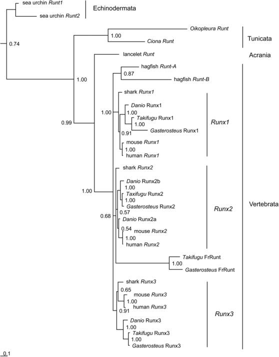
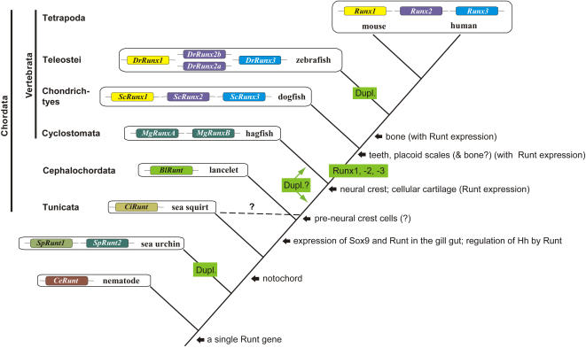
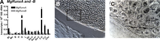
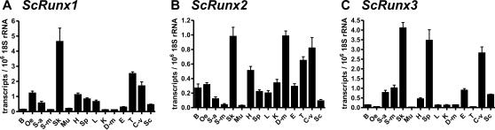
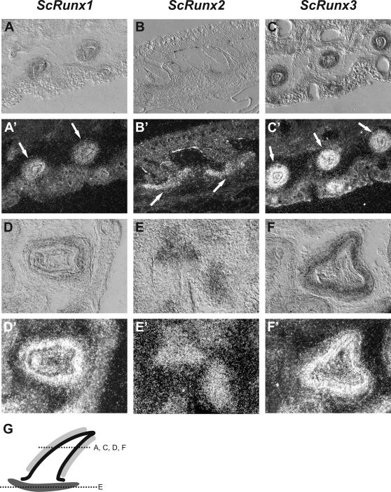
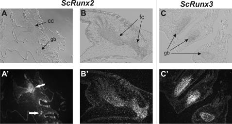
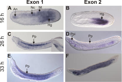
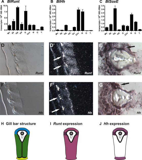
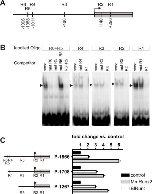
The skeleton is one of the most important features for the reconstruction of vertebrate phylogeny but few data are available to understand its molecular origin. In mammals the Runt genes are central regulators of skeletogenesis. Runx2 was shown to be essential for osteoblast differentiation, tooth development, and bone formation. Both Runx2 and Runx3 are essential for chondrocyte maturation. Furthermore, Runx2 directly regulates Indian hedgehog expression, a master coordinator of skeletal development. To clarify the correlation of Runt gene evolution and the emergence of cartilage and bone in vertebrates, we cloned the Runt genes from hagfish as representative of jawless fish (MgRunxA, MgRunxB) and from dogfish as representative of jawed cartilaginous fish (ScRunx1-3). According to our phylogenetic reconstruction the stem species of chordates harboured a single Runt gene and thereafter Runt locus duplications occurred during early vertebrate evolution. All newly isolated Runt genes were expressed in cartilage according to quantitative PCR. In situ hybridisation confirmed high MgRunxA expression in hard cartilage of hagfish. In dogfish ScRunx2 and ScRunx3 were expressed in embryonal cartilage whereas all three Runt genes were detected in teeth and placoid scales. In cephalochordates (lancelets) Runt, Hedgehog and SoxE were strongly expressed in the gill bars and expression of Runt and Hedgehog was found in endo- as well as ectodermal cells. Furthermore we demonstrate that the lancelet Runt protein binds to Runt binding sites in the lancelet Hedgehog promoter and regulates its activity. Together, these results suggest that Runt and Hedgehog were part of a core gene network for cartilage formation, which was already active in the gill bars of the common ancestor of cephalochordates and vertebrates and diversified after Runt duplications had occurred during vertebrate evolution. The similarities in expression patterns of Runt genes support the view that teeth and placoid scales evolved from a homologous developmental module.


Conflict of interest statement

The authors have declared that no competing interests exist.

Figures

Figure 1
Figure 1. Phylogenetic tree (Bayesian inference) of chordate Runt genes.
Numbers refer to branch support (Bayesian posterior probability) for the internal branches adjacent to the nodes. Sea urchin Runt genes were used to root the tree. Branch length reflects the number of substitutions per alignment site (compare scale bar).
Figure 2
Figure 2. Overview of the Runt gene evolution in chordates.
The stepwise evolution of cartilage and bone and the most likely time intervals of Runt gene duplications (Dup) are indicated. The position of tunicates is contentious which is indicated by a dashed line. In this context it is of interest that pre-neural crest cells have been observed in tunicates .
Figure 3
Figure 3. Analysis of hagfish MgRunxA and –B expression in different tissues of adult animals.
Quantification of MgRunxA and –B expression by qRT-PCR (A). Whereas MgRunxB was only weakly expressed in all tissues analyzed, MgRunxA showed a strong expression in calcified cartilage gills and soft cartilage. Expression of MgRunxA was also detected by radioactive in situ hybridisation in hard cartilage tissue (B, C). Insert of (B) is shown at higher magnification in (C) displaying the silver grains of the autoradiography emulsion indicating MgRunxA expression. B: Brain, C-h: Hard cartilage, C-s: Soft cartilage, Cho: Chorda, G: Gills, Gb: Gall bladder, G-a: Anterior gut, G-m: Midgut, G-h: Hindgut, H: Heart, L: Liver, Mu: Muscle, Sk: Skin.
Figure 4
Figure 4. qRT-PCR results of ScRunx1–3 expression.
In dogfish the most prominent expression of all three ScRunt genes was in the skin. Also in visceral cartilage ScRunx1–3 were strongly expressed. B: Brain, C-v: Visceral cartilage, D-m: Ductus mesonephric, E: Epididymis, H: Heart, K: Kidney, L: Liver, Mu: Muscle, Oe: Oesophagus, Sc: Spinal column, Sk: Skin, S-a: Anterior stomach, S-m: Middle part of stomach, Sp: Spleen, T: Testis.
Figure 5
Figure 5. ScRunx1–3 expression analysis by in situ hybridization in placoid scale (A–C) and tooth development (D–F).
Bright field is given on top, dark field below. ScRunx1 (A, D) and –3 (C, F) are expressed in the basal epidermis cells of the stratum germinativum, which forms the enamel organ, whereas ScRunx2 (B, E) is found at the site of the developing basal plate. These expression patterns were identical in teeth and placoid scales. (G) Scheme of Runt expression in placoid scales and teeth with overlapping expression of ScRunx1 and –3 in the stratum germinativum (light grey) and ScRunx2 in the developing basal plate (dark grey). Dotted lines represent section planes of transverse sections in (A,C–F). Section in (B) is a longitudinal section.
Figure 6
Figure 6. Expression of ScRunx2 and –3 in developing dogfish cartilage.
Expression of ScRunx2 was detected in developing cranial and gill bar cartilage (A) and in the proximal cartilage elements of the pectoral fin (B). Expression of ScRunx3 was detected in developing visceral cartilage (C). Cc: cranial cartilage, gb: gill gut cartilage, fc: fin cartilage.
Figure 7
Figure 7. Runt gene expression in lancelet larvae (B. floridae).
Anterior site is located to the left and the dorsal site towards the top. Whole mount in situ hybridization at stages of 16 h (A, B), 26 h (C, D) and 33 h (E, F). A), C) and E) Runt gene exon 1 variant. B), D) and F) Runt gene exon 2 variant. Note that the primary pigment spot, indicated by an arrow, lays in the nerve chord and does not represent a Runt expression domain. An: Anterior notochord, Nt: Neural tube, Nc: Notochord, Hg: Hindgut, Pp: Primary pigment spot, Ppi: Preoral pit.
Figure 8
Figure 8. Analysis of Runt, SoxE and Hh gene expression in adult lancelet (B. lanceolatum).
(A–C) Quantification of Runt, SoxE and Hh expression in different tissues. (A) The strongest Runt expression is seen in the gill gut region followed by the gut and skin. (B) Hh is most strongly expressed in the chorda and neural tube followed by the gill gut and gut. (C) SoxE has its strongest expression in the gill gut and neural tube. Mu: Muscle, Sk: Skin, Gg: Gill gut, Hd: Hepatic diverticulum, G: Gut, Cho: Chorda, N: Neural tube, O: Ovaries, T: Testis. (D–G) in situ hybridization for BlRunt and BlHh show high expression in the endoderm and ecotoderm of the gill bars. (D–E) Runt expression. (F–G) Hh expression. (D, F) Bright field images. (D′, F′) Dark field images of radioactive in situ hybridizations. (E, G) Non-radioactive in situ hybridizations. High expression of BlRunt and BlHh was found in a cell population between the endodermal epithelium with cilia and the ectodermal gland epithelium directly adjacent to both sites of the acellular matrix (arrows). (H–J) Schematic drawing of Runt and Hh expression sites in secondary gill bars as shown in (D–G). (H) The gill bar tissue consists of three different single layered epithelia attached to a basal membrane - atrial epithelium (blue), lateral epithelium (dark green) and pharyngeal epithelium (light green). The basal membrane is indicated by the bold black line. The skeletal rod of secondary gill bars contains a skeletal vessel (grey filled circle) that is formed by basal membranes, and does not contain endothelial cells. (I) Runt expression is found throughout the gill bar epithelia (light purple) with strongest expression adjacent to the skeletal rods (dark purple). (J) Hh is expressed at weaker levels in the atrial and pharyngeal epithelium (light purple) and at high levels in the cell population adjacent to the skeletal rods (dark purple).
Figure 9
Figure 9. Runt dependent regulation of the B. floridae Hh promoter.
(A) Scheme of the BfHh promoter with putative Runt binding sites. Number and position relative to the transcription start site is given. (B) Electrophoretic mobility shift assays using oligos containing R1–R6 Runt binding sites. BlRunt can bind to each of the putative binding sites. Strongest binding is observed for the oligo with the closely adjacent binding sites R5 and R6 and for the R1 oligo. Nuclear extracts without BlRunt do not show a mobility shift of the oligos (data not shown). (C) Runt dependent activation of the BfHh promoter in NIH3T3 cells. Overexpression of either BlRunt or MmRunx2 leads to activation of the indicated promoter constructs compared to constructs co-transfected with an empty expression vector.

References

    1. Hall BK. Bones and Cartilage, Developmental and Evolutionary Skeletal Biology. London: Elsevier Academic Press; 2005. p. 760.
    1. Robson P, Wright GM, Keeley FW. Distinct non-collagen based cartilages comprising the endoskeleton of the Atlantic hagfish, Myxine glutinosa. Anat Embryol (Berl) 2000;202:281–290. - PubMed
    1. Zhang G, Miyamoto MM, Cohn MJ. Lamprey type II collagen and Sox9 reveal an ancient origin of the vertebrate collagenous skeleton. Proc Natl Acad Sci U S A. 2006;103:3180–3185. - PMC - PubMed
    1. Zhang G, Cohn MJ. Hagfish and lancelet fibrillar collagens reveal that type II collagen-based cartilage evolved in stem vertebrates. Proc Natl Acad Sci U S A. 2006;103:16829–16833. - PMC - PubMed
    1. Rychel AL, Swalla BJ. Development and evolution of chordate cartilage. J Exp Zoolog B Mol Dev Evol. 2007;308:325–335. - PubMed

MeSH terms