. 2008 Mar 14;4(3):e1000036.
doi: 10.1371/journal.pgen.1000036.

Transcriptional profiling uncovers a network of cholesterol-responsive atherosclerosis target genes

Affiliations

Transcriptional profiling uncovers a network of cholesterol-responsive atherosclerosis target genes

Josefin Skogsberg et al. PLoS Genet. .

Abstract

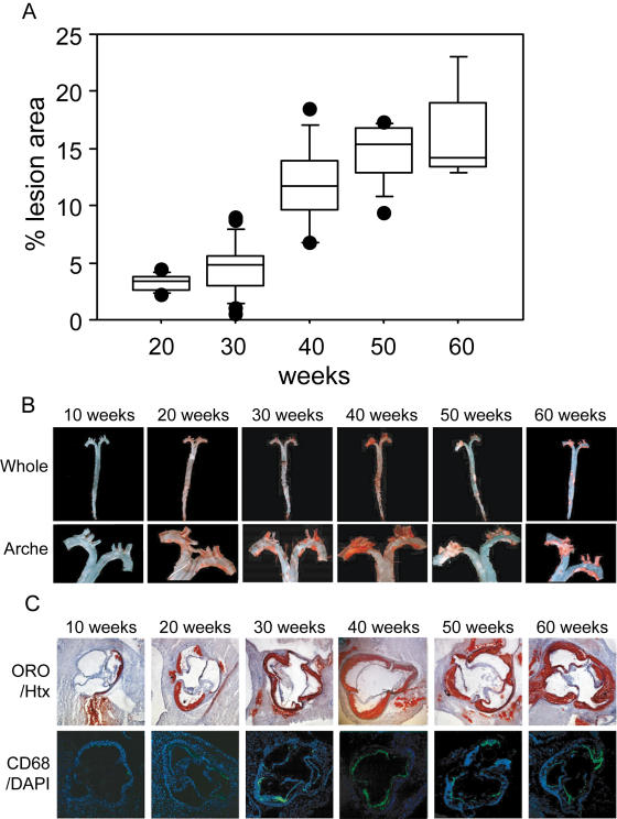
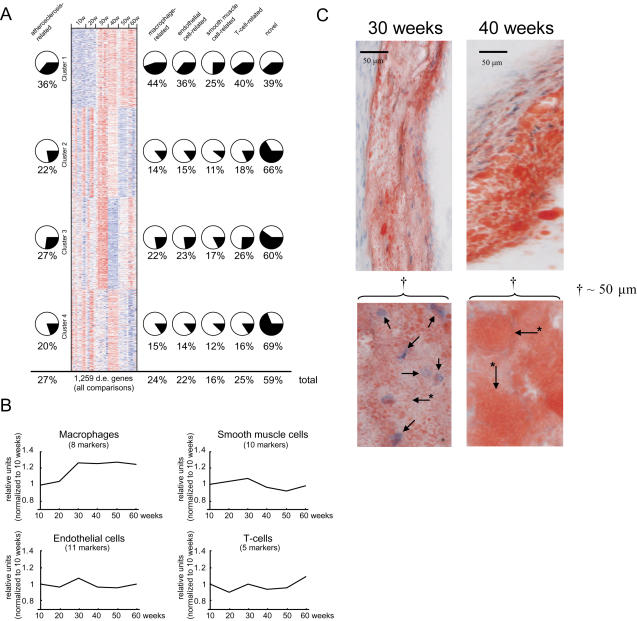
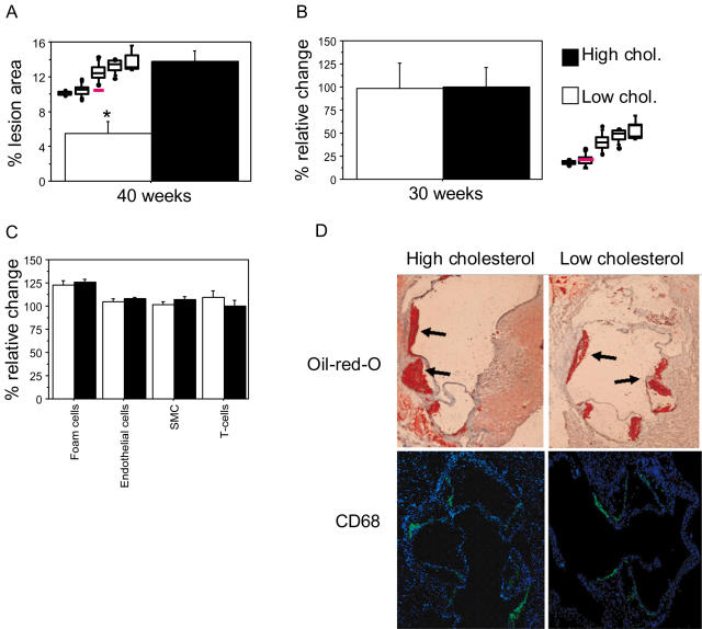
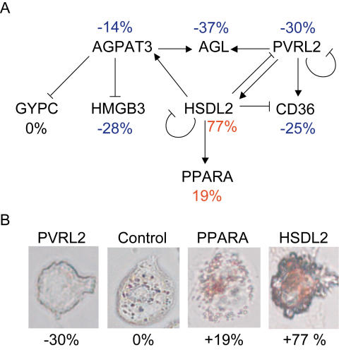
Despite the well-documented effects of plasma lipid lowering regimes halting atherosclerosis lesion development and reducing morbidity and mortality of coronary artery disease and stroke, the transcriptional response in the atherosclerotic lesion mediating these beneficial effects has not yet been carefully investigated. We performed transcriptional profiling at 10-week intervals in atherosclerosis-prone mice with human-like hypercholesterolemia and a genetic switch to lower plasma lipoproteins (Ldlr(-/-)Apo(100/100)Mttp(flox/flox) Mx1-Cre). Atherosclerotic lesions progressed slowly at first, then expanded rapidly, and plateaued after advanced lesions formed. Analysis of lesion expression profiles indicated that accumulation of lipid-poor macrophages reached a point that led to the rapid expansion phase with accelerated foam-cell formation and inflammation, an interpretation supported by lesion histology. Genetic lowering of plasma cholesterol (e.g., lipoproteins) at this point all together prevented the formation of advanced plaques and parallel transcriptional profiling of the atherosclerotic arterial wall identified 37 cholesterol-responsive genes mediating this effect. Validation by siRNA-inhibition in macrophages incubated with acetylated-LDL revealed a network of eight cholesterol-responsive atherosclerosis genes regulating cholesterol-ester accumulation. Taken together, we have identified a network of atherosclerosis genes that in response to plasma cholesterol-lowering prevents the formation of advanced plaques. This network should be of interest for the development of novel atherosclerosis therapies.


Conflict of interest statement

Johan Björkegren and Jesper Tegnér are stock owners in Clinical Gene Networks AB. The company has filed a provisional patent on the atherosclerosis gene network presented in this study.

Figures

Figure 1
Figure 1. Atherosclerosis progression in Ldlr −/− Apob100/100 Mttp flox/flox mice.
(A) Box plots of atherosclerosis progression at 20 (n = 12), 30 (n = 25), 40 (n = 15), 50 (n = 15), and 60 weeks (n = 10). P<0.05, 20 vs. 30 weeks; P<0.0001, 30 vs. 40 weeks; P<0.02, 40 vs. 50 weeks. Values are surface lesion areas assessed by Sudan IV staining of pinned-out aortas and given as a percentage of the surface of the entire aorta. Boxes enclose values between the 75th and 25th percentiles, bars indicate values between the 90th and 10th percentiles, and black dots indicate individual observations outside these boundaries. (B) Representative aortic trees (above) and arches (below). (C) Representative sections of the aortic root were stained with Oil-red-O and counterstained with hematoxylin (Htx) or show CD68 fluorescence and counterstained with DAPI.
Figure 2
Figure 2. Transcriptional changes of atherosclerosis development.
(A) Heat map of clustered mRNA levels (rows; red, high levels; blue, low levels) for genes differentially expressed (d.e.) in at least one pair-wise time-point comparison (FDR<0.05, n = 1259). Each column represents mRNA levels in one mouse (n = 5 to 7 per time point) at 10 to 60 weeks of age. Four gene clusters were identified by the cluster algorithm. Pie charts show percentages of genes related to atherosclerosis (Table S1) on the left and to the four atherosclerosis cell types (Tables S2, S3, S4, and S5) on the right. A substantial number of genes were related to more than one atherosclerosis cell type (Figure S1). The percentage of novel genes (i.e., not related to atherosclerosis or atherosclerosis cell types) in each cluster is shown on the far right. Overall percentages (total) are shown below. (B) Relative expression levels of cell-specific markers of atherosclerosis cell types (Table S7). The number of markers per cell type is indicated. The only statistically significant increase was in the number of foam cells, which increased by ∼30% between 20 and 30 weeks (P<0.001) and remained elevated at 60 weeks. (C) Representative sections of the aortic root (top) were stained with Oil-red-O (red in the figure) and counterstained with hematoxylin (Htx) (blue spots in the figure), bar in figure indicate 50 µm; higher-power views of 50 µm showing foam cells at 30 (arrows indicate macrophage Htx-stained nuclei, asterisk marked arrows point to the outer cell membrane) and 40 weeks (asterisk marked arrows point to the outer cell membrane of Oil-red-O-stained fat within macrophages, the nuclei are not visible) are shown below.
Figure 3
Figure 3. Effect of plasma-cholesterol lowering on lesion progression.
Lesion surface area was determined as the percentage of lesion area in relation to the total area of pinned-out aortas from the bifurcation to the aortic root. At 28 weeks of age, mice received intraperitoneal injections of pI-pC to induce recombination of Mttp in the liver and were sacrificed 12 weeks later or 1 week after cholesterol lowering had been achieved. High-cholesterol control mice were injected with PBS. (A) At 40 weeks of age, lesion surface area in mice with low plasma cholesterol (i.e., pI-pC-treated, n = 7) had not progressed and differed significantly from that in high-cholesterol controls at 40 weeks (i.e., PBS-treated, n = 6) (*P<0.005). Figure 1A is shown for comparison; red line indicates mice with low plasma cholesterol. (B–D) One week of low levels of cholesterol (30-week-old mice) did not affect lesion size (P = 0.96), (B) Shown are percent relative changes in lesion area. Figure 1A is shown for comparison; red line indicates the low-cholesterol group. (C) The numbers of foam cells (P = 0.52), endothelial cells (P = 0.49), smooth muscle cells (P = 0.18) (SMC), and T cells (P = 0.34) and (D) the staining of Oil-red-O (upper panels) and fluorescence of anti-rat antibodies binding to rat anti mouse CD68 (lower panels) in representative sections isolated from the aortic root of high-cholesterol (left panels) and low-cholesterol (right panels) mice. Arrows indicate Oil-red-O stained lesions.
Figure 4
Figure 4. A regulatory gene network of foam-cell formation.
Twelve cholesterol-responsive atherosclerosis genes (Table S12, in bold) were targeted in THP-1 macrophages using siRNA. Two days after transfection, siRNA-targeted macrophages and controls treated with nonspecific siRNA were incubated with AcLDL (50 µg/mL) for 48 hours; total RNA was isolated, and CE and lipid accumulation were determined. (A) Sixteen expression profiles (HG-U133_Plus_2 arrays, Affymetrix) from 12 siRNA experiments and four pooled controls were used to generate the regulatory gene network (Results) of 8 cholesterol-responsive genes involved in foam-cell formation, including PPARA and CD36. CE accumulation (given as average percentage next to each node) was decreased (blue) by siRNA inhibition of 5 genes and increased (red) by inhibition of 2 genes; inhibition of 1 gene had no effect (black) (see also Table 3). (B) Representative siRNA treated THP-1 cells after 48 incubation with AcLDL and stained with Oil-red-O.

References

    1. Ong HT. The statin studies: from targeting hypercholesterolaemia to targeting the high risk patient. Qjm. 2005;98:599–614. - PubMed
    1. Grines CL. The role of statins in reversing atherosclerosis: What the latest regression studies show. J Interv Cardiol. 2006;19:3–9. - PubMed
    1. Tuomisto TT, Binder BR, Yla-Herttuala S. Genetics, genomics and proteomics in atherosclerosis research. Ann Med. 2005;37:323–332. - PubMed
    1. Venter JC, Adams MD, Myers EW, Li PW, Mural RJ, et al. The sequence of the human genome. Science. 2001;291:1304–1351. - PubMed
    1. Lander ES, Linton LM, Birren B, Nusbaum C, Zody MC, et al. Initial sequencing and analysis of the human genome. Nature. 2001;409:860–921. - PubMed

Publication types

MeSH terms