. 2008 Mar 28;4(3):e1000042.
doi: 10.1371/journal.pgen.1000042.

ZIP4H (TEX11) deficiency in the mouse impairs meiotic double strand break repair and the regulation of crossing over

Affiliations

ZIP4H (TEX11) deficiency in the mouse impairs meiotic double strand break repair and the regulation of crossing over

Carrie A Adelman et al. PLoS Genet. .

Abstract

We have recently shown that hypomorphic Mre11 complex mouse mutants exhibit defects in the repair of meiotic double strand breaks (DSBs). This is associated with perturbation of synaptonemal complex morphogenesis, repair and regulation of crossover formation. To further assess the Mre11 complex's role in meiotic progression, we identified testis-specific NBS1-interacting proteins via two-hybrid screening in yeast. In this screen, Zip4h (Tex11), a male germ cell specific X-linked gene was isolated. Based on sequence and predicted structural similarity to the S. cerevisiae and A. thaliana Zip4 orthologs, ZIP4H appears to be the mammalian ortholog. In S. cerevisiae and A. thaliana, Zip4 is a meiosis-specific protein that regulates the level of meiotic crossovers, thus influencing homologous chromosome segregation in these organisms. As is true for hypomorphic Nbs1 (Nbs1(DeltaB/DeltaB)) mice, Zip4h(-/Y) mutant mice were fertile. Analysis of spermatocytes revealed a delay in meiotic double strand break repair and decreased crossover formation as inferred from DMC1 and MLH1 staining patterns, respectively. Achiasmate chromosomes at the first meiotic division were also observed in Zip4h(-/Y) mutants, consistent with the observed reduction in MLH1 focus formation. These results indicate that meiotic functions of Zip4 family members are conserved and support the view that the Mre11 complex and ZIP4H interact functionally during the execution of the meiotic program in mammals.


Conflict of interest statement

The authors have declared that no competing interests exist.

Figures

Figure 1
Figure 1. Mammalian Zip4h transcripts.
Alignment of Zip4h isoforms retrieved from public sequence databases indicate similar full-length isoforms are present from human and mouse testis extracts (A); accession #'s NM_031276 and NM_031384, respectively. A second human isoform isolated from testis contains a novel exon inserted between exons 2 and 3 of the full-length isoform (B); accession # NM_001003811. A C-terminally truncated transcript with an alternate terminal exon spliced to exon 24 was found in mouse ES cells (C); accession # AK082794. An N-terminally truncated isoform containing a novel 5′ untranslated exon spliced to exon 13 was isolated from human testis (D); accession # AK057391. Exons corresponding to ZIP4H fragment isolated in the yeast two hybrid screen (NBS1) and the antigen used for antibody generation (Antigen) are illustrated in A.
Figure 2
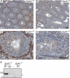
Figure 2. Zip4h expression in testis sections.
Testis sections from adult Zip4h+/Y (A, C) and Zip4h−/Y (B, D) mice were immunohistochemically labeled for ZIP4H (in brown), and counterstained with hematoxylin (in blue). Staining patterns under low magnification (A, B). Higher magnification images showing positively stained zygotene cells of control tubules (C) and absence of staining in mutant tubules at the same stage (D). Black bars represent 50 µm. ZIP4H IP-Western blot from Zip4h+/Y and Zip4h−/Y 12 d.p.p. testis extracts (E), asterisk denotes lower molecular weight species visible only in Zip4h−/Y samples.
Figure 3
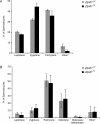
Figure 3. Meiotic stage distribution in mutant and control spermatocytes.
Spermatocyte spreads from 12 d.p.p. (A) and adult mice (B) were stained with SCP3 and SCP1 to analyze SC morphogenesis, and categorized according to meiotic stage. *Other denotes extensively fragmented or otherwise unidentifiable cells.
Figure 4
Figure 4. Persistent DSB repair intermediates in pachytene cells of Zip4h−/Y mice.
Spermatocyte spreads prepared from adult Zip4h+/Y and Zip4h−/Y mice were stained for DMC1 (green) and SCP3 (red). Representative images of pachytene stage spermatocytes from wildtype (A, B) and mutant (C, D) mice are shown. XY bivalents are indicated by arrowheads; asterisk in B indicates XY bivalent from adjacent spread.
Figure 5
Figure 5. Zip4h−/Y mice exhibit perturbations with respect to crossover formation.
Numbers of MLH1 foci per cell were tabulated in MLH1 positive spermatocyte spreads from Zip4h+/Y and Zip4−/Y mice and percent spreads containing the indicated foci were graphed (A). To determine whether DSB repair foci persisted to the stage when crossovers form, cells were co-stained with antibodies against MLH1 (red), RAD51 (green) and SCP3 (blue). Representative bivalents from Zip4h+/Y (B), and Zip4h−/Y (C) spreads are shown.
Figure 6
Figure 6. Crossover interference is reduced in Zip4h mutants.
SC lengths (A) and inter-crossover distance expressed as % of SC (B) of double exchange bivalents in Zip4h+/Y and Zip4h−/Y spermatocytes are graphed according to increasing length. Maximum likelihood fitting of the data to the gamma-distribution allows estimation of the interference parameter, K, for control (C) and mutant (D) double exchange bivalents.
Figure 7
Figure 7. Increased achiasmate chromosomes in MI cells from Zip4h−/Y mice.
Metaphase I spindles from stage XII tubules of Zip4h+/Y (A) and Zip4h−/Y (B) sections. Arrow indicates laggard. Representative diakinesis spreads from a wildtype mouse with no achiasmate chromosomes (C), and a mutant mouse exhibiting achiasmate XY chromosomes (D).
Figure 8
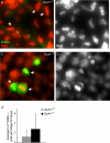
Figure 8. TUNEL positive metaphase cells in testis of Zip4h−/Y mice.
Testis sections from mutant and control mice were TUNEL labeled (green) and counterstained with DAPI (pseudocolored red) to assess cell death. TUNEL negative stage XII section from a wildtype mouse (A, B) containing cells with metaphase configurations (A, arrows; same image in B showing only greyscale DAPI). TUNEL positive cells from a Zip4h−/Y mouse (C, D), some exhibiting metaphase configurations (arrows). Average numbers of TUNEL positive cells per stage XII tubule are graphed with standard deviations (E).

References

    1. Richardson C, Horikoshi N, Pandita TK. The role of the DNA double-strand break response network in meiosis. DNA Repair (Amst) 2004;3:1149–1164. - PubMed
    1. Petronczki M, Siomos MF, Nasmyth K. Un menage a quatre: the molecular biology of chromosome segregation in meiosis. Cell. 2003;112:423–440. - PubMed
    1. Zickler D, Kleckner N. Meiotic chromosomes: integrating structure and function. Annu Rev Genet. 1999;33:603–754. - PubMed
    1. Eaker S, Cobb J, Pyle A, Handel MA. Meiotic prophase abnormalities and metaphase cell death in MLH1-deficient mouse spermatocytes: insights into regulation of spermatogenic progress. Dev Biol. 2002;249:85–95. - PubMed
    1. Hodges CA, Revenkova E, Jessberger R, Hassold TJ, Hunt PA. SMC1beta-deficient female mice provide evidence that cohesins are a missing link in age-related nondisjunction. Nat Genet. 2005;37:1351–1355. - PubMed

Publication types

MeSH terms