Review
. 2008 Nov;35(4):719-30.
doi: 10.1007/s00726-008-0062-5. Epub 2008 Mar 28.

Structural biology of proline catabolism

Affiliations
Review

Structural biology of proline catabolism

John J Tanner. Amino Acids. 2008 Nov.

Abstract

The proline catabolic enzymes proline dehydrogenase and Delta(1)-pyrroline-5-carboxylate dehydrogenase catalyze the 4-electron oxidation of proline to glutamate. These enzymes play important roles in cellular redox control, superoxide generation, apoptosis and cancer. In some bacteria, the two enzymes are fused into the bifunctional enzyme, proline utilization A. Here we review the three-dimensional structural information that is currently available for proline catabolic enzymes. Crystal structures have been determined for bacterial monofunctional proline dehydrogenase and Delta(1)-pyrroline-5-carboxylate dehydrogenase, as well as the proline dehydrogenase and DNA-binding domains of proline utilization A. Some of the functional insights provided by analyses of these structures are discussed, including substrate recognition, catalytic mechanism, biochemical basis of inherited proline catabolic disorders and DNA recognition by proline utilization A.


Figures
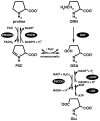
Fig. 1
Fig. 1
Reactions catalyzed by proline catabolic and biosynthetic enzymes.
Fig. 2
Fig. 2
Unrooted phylogenetic tree representing the organization of proline catabolic enzymes in bacteria and eukaryotes. The tree has three main branches corresponding to PutAs (branches 1 and 2) and monofunctional enzymes (branch 3). PutAs are found only in bacteria. The monofunctional enzymes appear in both eukaryotes (branch 3A) and bacteria (branch 3B). Structures of proline catabolic proteins and domains are superimposed on their respective branches (clockwise from top left): E. coli PutA DNA-binding domain (branch 1), E. coli PutA PRODH domain (branch 1), T. thermophilus TtPRODH (branch 3B) and T. thermophilus TtP5CDH (branch 3B).
Fig. 3
Fig. 3
Two views of the monofunctional PRODH from T. thermophilus. β-strands and α-helices of the (βα)8 barrel are colored magenta and cyan, respectively. Helix 8 is colored red. Selected strands and helices are numbered. The FAD cofactor is shown in yellow sticks. This figure and others were prepared with PyMOL (W. L. DeLano (2002) The PyMOL Molecular Graphics System).
Fig. 4
Fig. 4
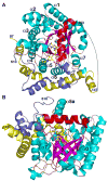
Two views of the PRODH domain of E. coli PutA complexed with the proline analog THFA. The orientations shown here are similar to those of Fig. 3. β-strands and α-helices of the (βα)8 barrel are colored magenta and cyan, respectively. Helix 8 is colored red. Selected strands and helices are numbered. The FAD cofactor and THFA inhibitor are shown in yellow and green sticks, respectively. The helical elbow that wraps around the barrel is shown in yellow. Residues following the barrel are colored slate. The dotted curve indicates poorly ordered parts of the polypeptide chain between residues 147 and 263.
Fig. 5
Fig. 5
Stereo view of the active site the E. coli PutA PRODH domain highlighting interactions with the inhibitor THFA (green). The FAD cofactor is colored yellow. Protein side chains are colored white. Dotted lines indicate hydrogen bonds and ion pairs.
Fig. 6
Fig. 6
Schematic diagram of interactions between the E. coli PutA PRODH domain and the proline analog THFA. Dotted lines indicate hydrogen bonds and ion pairs. Thick solid lines indicate van der Waals interactions.
Fig. 7
Fig. 7
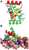
Structure of the E. coli PutA DNA-binding domain. A, PutA52 dimer highlighting residues important for binding DNA. The two chains are colored green and red. Positions of residues of the β-sheet that form hydrogen bonds with DNA bases are indicated in yellow. Side chains of residues at the N-terminus of αB that interact with the DNA backbone are drawn as cyan sticks. B, Structure of PutA52 complexed with DNA, highlighting interaction of the protein β-sheet with the DNA major groove. Coloring of the protein is the same as in panel A. Selected protein side chains are drawn as spheres.
Fig. 8
Fig. 8
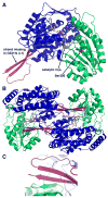
Structure P5CDH from T. thermophilus (Inagaki et al. 2006). A, ribbon drawing of a P5CDH subunit. The three domains are colored blue (NAD+-binding), green (catalytic) and pink (dimerization). The NAD+ cofactor is shown in yellow sticks. Catalytic Cys322 is represented in spheres. B, ribbon drawing of a P5CDH dimer. The two subunits of the dimer are colored as in panel A. C, close-up view of the intermolecular β-sheet in the dimer interface.
Scheme 1
Scheme 1

Similar articles

Cited by

References

    1. Adams E, Frank L. Metabolism of proline and the hydroxyprolines. Annu Rev Biochem. 1980;49:1005–1061. - PubMed
    1. Baban BA, Vinod MP, Tanner JJ, Becker DF. Probing a hydrogen bond pair and the FAD redox properties in the proline dehydrogenase domain of Escherichia coli PutA. Biochim Biophys Acta. 2004;1701:49–59. - PubMed
    1. Bearne SL, Wolfenden R. Glutamate gamma-semialdehyde as a natural transition state analogue inhibitor of Escherichia coli glucosamine-6-phosphate synthase. Biochemistry. 1995;34:11515–11520. - PubMed
    1. Becker DF, Thomas EA. Redox properties of the PutA protein from Escherichia coli and the influence of the flavin redox state on PutA-DNA interactions. Biochemistry. 2001;40:4714–4721. - PubMed
    1. Bender HU, Almashanu S, Steel G, Hu CA, Lin WW, Willis A, Pulver A, Valle D. Functional consequences of PRODH missense mutations. Am J Hum Genet. 2005;76:409–420. - PMC - PubMed

Publication types

LinkOut - more resources