Review
. 2008:450:3-26.
doi: 10.1007/978-1-60327-214-8_1.

The development of germline stem cells in Drosophila

Affiliations
Review

The development of germline stem cells in Drosophila

David A Dansereau et al. Methods Mol Biol. 2008.

Abstract

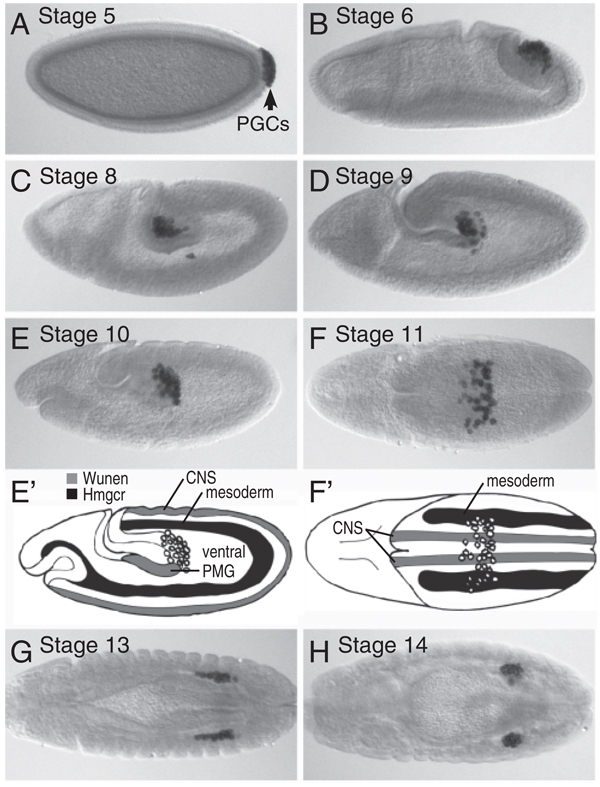
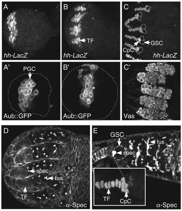
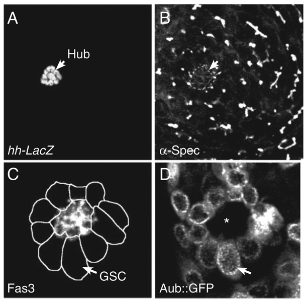
Germline stem cells (GSCs) in Drosophila are a valuable model to explore of how adult stem cells are regulated in vivo. Genetic dissection of this system has shown that stem cell fate is determined and maintained by the stem cell's somatic microenvironment or niche. In Drosophila gonads, the stem cell niche -- the cap cell cluster in females and the hub in males -- acts as a signaling center to recruit GSCs from among a small population of undifferentiated primordial germ cells (PGCs). Short-range signals from the niche specify and regulate stem cell fate by maintaining the undifferentiated state of the PGCs next to the niche. Germline cells that do not receive the niche signals because of their location assume the default fate and differentiate. Once GSCs are specified, adherens junctions maintain close association between the stem cells and their niche and help to orient stem cell division so that one daughter is displaced from the niche and differentiates. In females, stem cell fate depends on bone morphogenetic protein (BMP) signals from the cap cells; in males, hub cells express the cytokine-like ligand Unpaired, which activates the Janus kinase-signal transducers and activators of transcription (Jak-Stat) pathway in stem cells. Although the signaling pathways operating between the niche and stem cells are different, there are common general features in both males and females, including the arrangement of cell types, many of the genes used, and the logic of the system that maintains stem cell fate.


Figures

Fig. 1.1
Fig. 1.1
Primordial germ cell migration. Lateral view, dorsal up, anterior left (A–E). The germ-line is the first distinct cell lineage established in the Drosophila embryo when primordial germ cells (PGCs), or pole cells, cellularize at the posterior pole of the embryo (A). PGC fate is determined by their inheritance of polar granules, which are located in the posterior cytoplasm of the embryo. A polar granule component, Vasa, is used here to mark the germline (A–H). PGCs are carried dorsally and anteriorly as the germ band extends, and during gastrulation, the posterior midgut (PMG) invaginates and carries the PGCs into the PMG lumen (B and C). PGCs then migrate through the epithelium of the PMG into the interior of the embryo (D and E). PGCs migrate away from Wunen expression in the ventral PMG (grey in E') and toward the 3-hydroxy-3-methylglutaryl coenzyme A (HMG-CoA) reductase (Hmgcr)-expressing mesoderm (black in E') and thus move dorsally. Dorsal view, anterior left (F–H). PGCs migrate toward the Hmgcrexpressing mesoderm (black in F') and split into two groups as they are repelled from the midline by Wunen expression in the central nervous system (CNS; gray in F'). Within the mesoderm, PGCs associate with the somatic gonadal precursors (SGPs) in parasegments 10–12 (G), before migrating anteriorly and coalescing to form embryonic gonads in parasegment 10 (H)
Fig. 1.2
Fig. 1.2
Female germline stem cell niche development. hh-LacZ is specifically expressed in the developing GSC niche in the anterior of the developing ovary (A–C). Niche development commences in early third larval instar with the identity of somatic terminal filament (TF) cells in the anterior of the gonad. Terminal filament cells intercalate and separate into clusters (A), then extend by cell shape changes and by cell recruitment during late third larval instar (B). The niche develops anterior to the primordial germ cell (PGC) population, which is located in the medial region of the ovary, and contacts a subset of PGCs, marked here with Aub∷GFP (A', B'). Cap cells (CpC) are formed in pupal ovaries after TF formation is complete (C). GSC fate is restricted to anterior PGCs during the early pupal stage based on their juxtaposition to cap cells (D, early pupae; E, late pupae). Antibodies to α-Spectrin mark the cell membranes of TF cells, the spectrosomes of PGCs and GSCs (spec), and the fusomes of differentiating germline cells (fus). Anterior PGCs next to the developing niche maintain a single spectrosome that becomes attached to the CpC–GSC contact site. The differentiation of germline cells further away from the niche is marked by the presence of branched fusomes (D, E)
Fig. 1.3
Fig. 1.3
The germline stem cell (GSC) niche of male third instar larvae. GSCs in the testis are maintained by a cluster of somatic hub cells that specifically express an hh-lacZ enhancer trap (A) and Fas3 protein (C). The position of the hub from A is noted in B (arrow). Antibodies to α-Spectrin mark GSC spectrosomes next to the hub and the fusomes of differentiating gonialblasts and dividing cysts (B). GSCs are arranged around the hub in a characteristic rosette pattern (C). The positions of GSCs from D are noted around the Fas3-expressing hub in C. Germline cells arranged around the hub (*) express an Aub∷GFP (green fluorescent protein) fusion protein (D)

Similar articles

Cited by

References

    1. Fuchs E, Tumbar T, Guasch G. Socializing with the neighbors: stem cells and their niche. Cell. 2004;116:769–778. - PubMed
    1. Wong MD, Jin Z, Xie T. Molecular mechanisms of germline stem cell regulation. Annu. Rev. Genet. 2005;39:173–195. - PubMed
    1. Saffman EE, Lasko P. Germline development in vertebrates and invertebrates. Cell Mol. Life Sci. 1999;55:1141–1163. - PMC - PubMed
    1. Mahowald AP. Assembly of the Drosophila germ plasm. Int. Rev. Cytol. 2001;203:187–213. - PubMed
    1. Seydoux G, Schedl T. The germline in C. elegans: origins, proliferation, and silencing. Int. Rev. Cytol. 2001;203:139–185. - PubMed

Publication types

MeSH terms

Substances

LinkOut - more resources