. 2008:439:147-58.
doi: 10.1007/978-1-59745-188-8_10.

Transcriptional profiling of small samples in the central nervous system

Affiliations

Transcriptional profiling of small samples in the central nervous system

Stephen D Ginsberg. Methods Mol Biol. 2008.

Abstract

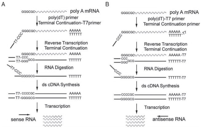
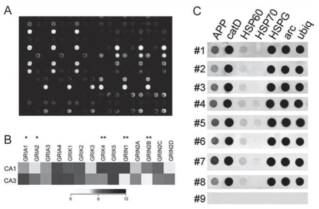
RNA amplification is a series of molecular manipulations designed to amplify genetic signals from small quantities of starting materials (including single cells and homogeneous populations of individual cell types) for microarray analysis and other downstream genetic methodologies. A novel methodology named terminal continuation (TC) RNA amplification has been developed in this laboratory to amplify RNA from minute amounts of starting material. Briefly, an RNA synthesis promoter is attached to the 3' and/or 5' region of cDNA utilizing the TC mechanism. The orientation of amplified RNAs is "antisense" or a novel "sense" orientation. TC RNA amplification is utilized for many downstream applications, including gene expression profiling, microarray analysis, and cDNA library/subtraction library construction. Input sources of RNA can originate from a myriad of in vivo and in vitro tissue sources. Moreover, a variety of fixations can be employed, and tissues can be processed for histochemistry or immunocytochemistry prior to microdissection for TC RNA amplification, allowing for tremendous cell type and tissue specificity of downstream genetic applications.


Figures

Fig. 10.1
Fig. 10.1
Overview and analysis of the TC RNA amplification method. (A) A TC primer (containing a bacteriophage promoter sequence for sense orientation) and a poly d(T) primer are added to the mRNA population to be amplified. First-strand synthesis occurs as an mRNA-cDNA hybrid is formed after reverse transcription and terminal continuation of the oligonucleotide primers. Following RNase H digestion to remove the original mRNA template strand, second-strand synthesis is performed using Taq polymerase. The resultant double-stranded product is utilized as template for IVT, yielding high-fidelity, linear RNA amplification of sense orientation (rippled lines). (B) Schematic similar to A, illustrating the TC RNA amplification procedure amplifying RNA in the antisense orientation (rippled lines). Adapted from Ginsberg 2005 [26]
Fig. 10.2
Fig. 10.2
Representative array platforms illustrating utility of RNA amplification procedures for single cell and population cell analysis. (A) Spotted cDNA array platform using RNA amplified from individual hippocampal CA1 pyramidal neurons from normal control brains and from neurofibrillary tangle (NFT) bearing CA1 neurons from Alzheimer’s disease (AD) patients. (B) Dendrogram demonstrating relative expression levels of representative genes in CA1 and CA3 pyramidal neurons microaspirated from human hippocampus. A heatmap matrix plot illustrates relative expression levels for individual glutamate receptor transcripts in CA1 and CA3 neurons. A single asterisk indicates a significant increase in expression in CA3 neurons as compared to CA1 neurons and a double asterisk denotes a significant increase in expression in CA1 neurons as compared to CA3 neurons. Key: GRIA 1, alpha-amino-3-hydroxy-5-methyl-4-isoxa-zolepropionate receptor 1 (AMPA1); GRIA2, AMPA2, GRIA3, AMPA3, GRIA4, AMPA4; GRIK1, kainate (KA) receptor GluR5; GRIK2, KA GluR6; GRIK3, KA GluR7; GRIK4, KA receptor KA1; GRIK5, KA receptor KA2; GRIN1, N-methyl D-aspartate receptor 1 (NR1); GRIN2A, NR2A; GRIN2B, NR2B; GRIN2C, NR2C; GRIN2D, NR2D. Adapted from Ginsberg and Che 2005 [32]. (C) Single-cell array analysis of human CA1 pyramidal neurons using custom-designed cDNA arrays and TC RNA amplification. Representative arrays illustrate a wide dynamic range of hybridization signal intensities for eight human postmortem cases (numbers 1–8). The negative control (number 9) is a single CA1 pyramidal neuron from the first case (number 1) that does not have the primers necessary for TC RNA amplification. In addition, a moderate variation of gene level expression across the eight human cases is observed, indicating the utility of using postmortem human samples for normative and neuropathological investigations. Key: APP, amyloid-β precursor protein; catD, cathepsin D; HSP60, heat shock protein 60; HSP70, heat shock protein 70; arc, activity regulated cytoskeletal-associated protein; ubiq, ubiquitin thiolesterase. Adapted from Ginsberg and Che 2002 [10]

References

    1. Ginsberg SD, Mirnics K. Functional genomic methodologies. Prog Brain Res. 2006;158:15–40. - PubMed
    1. Kabbarah O, et al. Expression profiling of mouse endometrial cancers microdissected from ethanol-fixed, paraffin-embedded tissues. Am J Pathol. 2003;162:755–762. - PMC - PubMed
    1. Su JM, et al. Comparison of ethanol versus formalin fixation on preservation of histology and RNA in laser capture microdissected brain tissues. Brain Pathol. 2004;14:175–182. - PMC - PubMed
    1. Tanji N, et al. Effect of tissue processing on the ability to recover nucleic acid from specific renal tissue compartments by laser capture microdissection. Exp Nephrol. 2001;9:229–234. - PubMed
    1. Fend F, et al. Immuno-LCM: Laser capture microdissection of immunostained frozen sections for mRNA analysis. Am J Pathol. 1999;154:61–66. - PMC - PubMed

Publication types

LinkOut - more resources