. 2008 Apr 22;47(16):4614-20.
doi: 10.1021/bi702069d. Epub 2008 Mar 28.

Spectral tuning of deep red cone pigments

Affiliations

Spectral tuning of deep red cone pigments

Tabitha L Amora et al. Biochemistry. .

Abstract

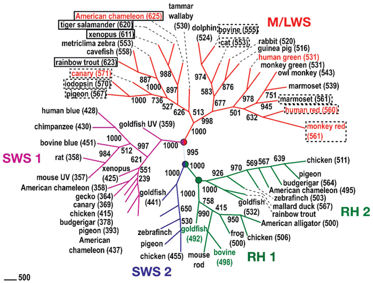
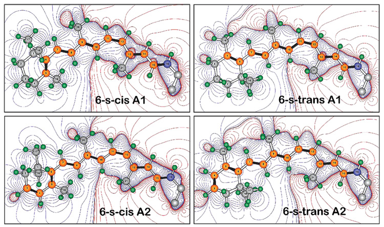
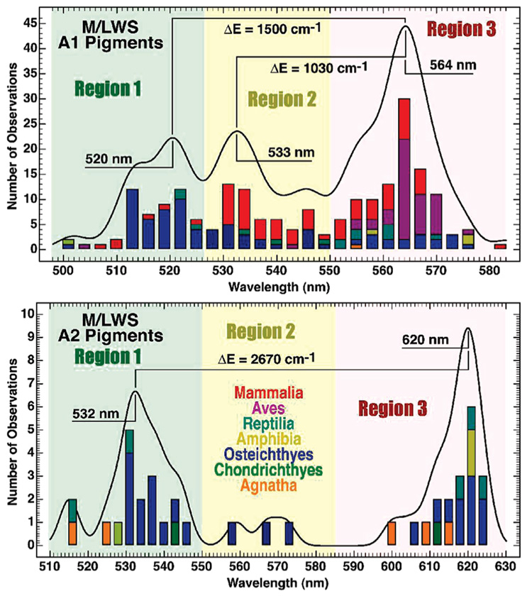
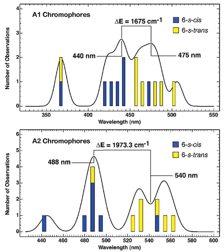
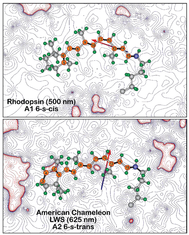
Visual pigments are G-protein-coupled receptors that provide a critical interface between organisms and their external environment. Natural selection has generated vertebrate pigments that absorb light from the far-UV (360 nm) to the deep red (630 nm) while using a single chromophore, in either the A1 (11- cis-retinal) or A2 (11- cis-3,4-dehydroretinal) form. The fact that a single chromophore can be manipulated to have an absorption maximum across such an extended spectral region is remarkable. The mechanisms of wavelength regulation remain to be fully revealed, and one of the least well-understood mechanisms is that associated with the deep red pigments. We investigate theoretically the hypothesis that deep red cone pigments select a 6- s- trans conformation of the retinal chromophore ring geometry. This conformation is in contrast to the 6- s- cis ring geometry observed in rhodopsin and, through model chromophore studies, the vast majority of visual pigments. Nomographic spectral analysis of 294 A1 and A2 cone pigment literature absorption maxima indicates that the selection of a 6- s- trans geometry red shifts M/LWS A1 pigments by approximately 1500 cm (-1) ( approximately 50 nm) and A2 pigments by approximately 2700 cm (-1) ( approximately 100 nm). The homology models of seven cone pigments indicate that the deep red cone pigments select 6- s- trans chromophore conformations primarily via electrostatic steering. Our results reveal that the generation of a 6- s- trans conformation not only achieves a significant red shift but also provides enhanced stability of the chromophore within the deep red cone pigment binding sites.


Figures

FIGURE 1
FIGURE 1
This study’s premise is that many of the deep red cones are bathochromically shifted to higher wavelength by adopting a 6-s-trans conformation of the 11-cis-retinal chromophore. The figure places solid rectangles around those pigments for which A2 6-s-trans conformations are predicted, with dashed lines indicating more tentative assignments for the A1 6-s-trans pigments. The phylogenetic tree shown here is constructed from aligned sequences by neighbor-joining methods through ClustalW. The distances of the sequences are estimated on the basis of the Dayhoff PAM matrix by Prodist of the PHYLIP program (version 3.65). The numbers along the branches indicate the clustering percentage obtained from 1000 bootstrap resamplings. The values enclosed in parentheses are the observed absorption maxima of the pigments, when available. Classification nodes are marked with circles, and the seven pigments modeled in this study are labeled in color.
FIGURE 2
FIGURE 2
Vertebrate visual pigments select either A1 retinal or A2 (3,4-dehydroretinal) 11-cis-retinal as the bound chromophore. A2 retinal has an additional double bond in the β-ionone ring, which red shifts the absorption spectrum. The contours describe the differential electrostatic field based on the point-charge Mulliken population [B3LYP/6-31G(d)]. The red contours indicate areas of positive charge, and the blue contours indicate areas of negative charge relative to the chromophore as a whole, which carries a net positive charge. Note that the 6-s-trans conformers move positive charges toward the ring relative to the 6-s-cis conformers.
FIGURE 3
FIGURE 3
Histogram analysis of 294 M/LWS pigments reveals the distribution of absorption maxima into two or three regions of the visible spectrum for A1 (top) and A2 (bottom) pigments. We refer to those red cones with absorption maxima in region 3 as “deep red” cones and propose that these pigments have 6-s-trans chromophores. The literature data and associated references are listed in Tables S1 and S2 of the Supporting Information.
FIGURE 4
FIGURE 4
Histogram of the transition energies of the 10 model chromophores calculated using the MNDO-PSDCI theory including full single- and double-configuration interaction within the π system. The structures and the data are listed in Tables S3 and S4 in the Supporting Information. The calculations confirm that the 6-s-trans conformers are on average red-shifted relative to the 6-s-cis conformers. A histogram analysis reveals that the difference in the transition wavelength between the 6-s-trans and the 6-s-cis conformers is similar to that observed between regions 1 and 3 of the experimental A1 and A2 histograms (Figure 3).
FIGURE 5
FIGURE 5
Electrostatic contours in the plane of the chromophores of bovine rhodopsin (1U19) and a 6-s-trans homology model of American chameleon based on 1U19. The contours are based on Mulliken charges from a single SCF PM3 Mozyme calculation on a Charmm-based structure following 2 ns molecular dynamics. The contours are associated with the protein residues, ignoring the chromophore charges. Note that, in both cases, the chromophore is bathed in an electrostatic field that is predominantly negative (blue contours). However, the dipole moment of the binding site is nearly orthogonal between the two proteins, with the rhodopsin binding site favoring a 6-s-cis conformer (top) and the American chameleon binding site (bottom) favoring a 6-s-trans conformer.

References

    1. Ebrey T, Koutalos Y. Vertebrate photoreceptors. Prog. Retinal Eye Res. 2001;20:49–94. - PubMed
    1. Yokoyama S, Radlwimmer FB. The molecular genetics and evolution of red and green color vision in vertebrates. Genetics. 2001;158:1697–1710. - PMC - PubMed
    1. Dartnall HJ, Lythgoe JN. The spectral clustering of visual pigments. Vision Res. 1965;5:81–100. - PubMed
    1. Harosi FI. An analysis of two spectral properties of vertebrate visual pigments. Vision Res. 1994;34:1359–1367. - PubMed
    1. Reckel F, Melzer RR, Parry JWL, Bowmaker JK. The retina of five atherinomorph teleosts: Photoreceptors, patterns and spectral sensitivities. Brain, Behav., Evol. 2002;60:249–264. - PubMed

Publication types