

The evolution of our understanding on glioma
- PMID: 18371180
- PMCID: PMC8095536
- DOI: 10.1111/j.1750-3639.2008.00136.x
The evolution of our understanding on glioma
Abstract
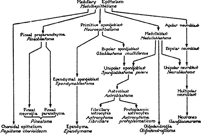
The description of neuroglia by Virchow in 1848 may be considered the starting point of our understanding of primary brain tumors. At the beginning of the 20th century, surgical removal of primary brain tumors became possible, and therefore, tissue for microscopic analysis and clinical data on survival became available. During this time, research on gliomas beyond improving surgical procedures focused on their classification. The classification schemes developed emphasized parameters for sorting tumors with regard to (i) cytological aspects; (ii) presumed tumor cell origin; (iii) histological appearance of the tissue; or (iv) clinical outcome. Over the years, experimental studies have greatly improved our knowledge on gliomas. Gliomas induced by viruses, chemicals, radiation, transgenes and knock-out technology contributed to the understanding of their pathogenesis and still serve as preclinical models for the testing of novel therapies. Recent advances in developmental neurobiology and the identification of stem cells provided new insights into the origin of brain tumors and the molecular mechanisms of tumor formation. This review briefly compiles the evolution of our concepts on gliomas, focusing on the latest developments.
Figures
References
-
- Aguzzi A, Kleihues P, Heckl K, Wiestler OD (1991) Cell type‐specific tumour induction in neural transplants by retrovirus‐mediated oncogene transfer. Oncogene 6:113–118. - PubMed
-
- Altman J, Das GD (1965) Post‐natal origin of microneurones in the rat brain. Nature 207:953–956. - PubMed
-
- Alvarez‐Buylla A, Garcia‐Verdugo JM, Tramontin AD (2001) A unified hypothesis on the lineage of neural stem cells. Nat Rev 2:287–293. - PubMed
-
- Bachoo RM, Maher EA, Ligon KL, Sharpless NE, Chan SS, You MJ et al (2002) Epidermal growth factor receptor and Ink4a/Arf: convergent mechanisms governing terminal differentiation and transformation along the neural stem cell to astrocyte axis. Cancer Cell 1:269–277. - PubMed
-
- Bailey P, Cushing H (1926) A Classification of The Tumors of The Glioma Group on a Histogenetic Basis with a Correlated Study of Prognosis. Lippincot JB: Philadelphia.
Publication types
MeSH terms
LinkOut - more resources
Full Text Sources
Other Literature Sources
Medical
