. 2008 Jul;18(3):370-7.
doi: 10.1111/j.1750-3639.2008.00130.x. Epub 2008 Mar 26.

CD133 expression and cancer stem cells predict prognosis in high-grade oligodendroglial tumors

Affiliations

CD133 expression and cancer stem cells predict prognosis in high-grade oligodendroglial tumors

Dagmar Beier et al. Brain Pathol. 2008 Jul.

Abstract

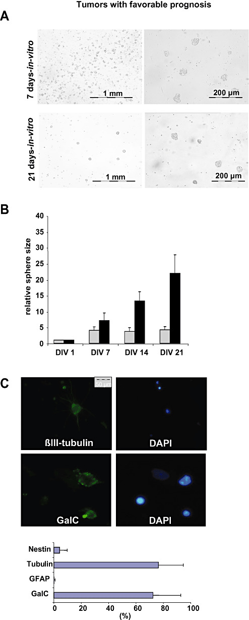
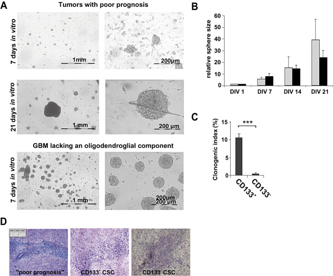
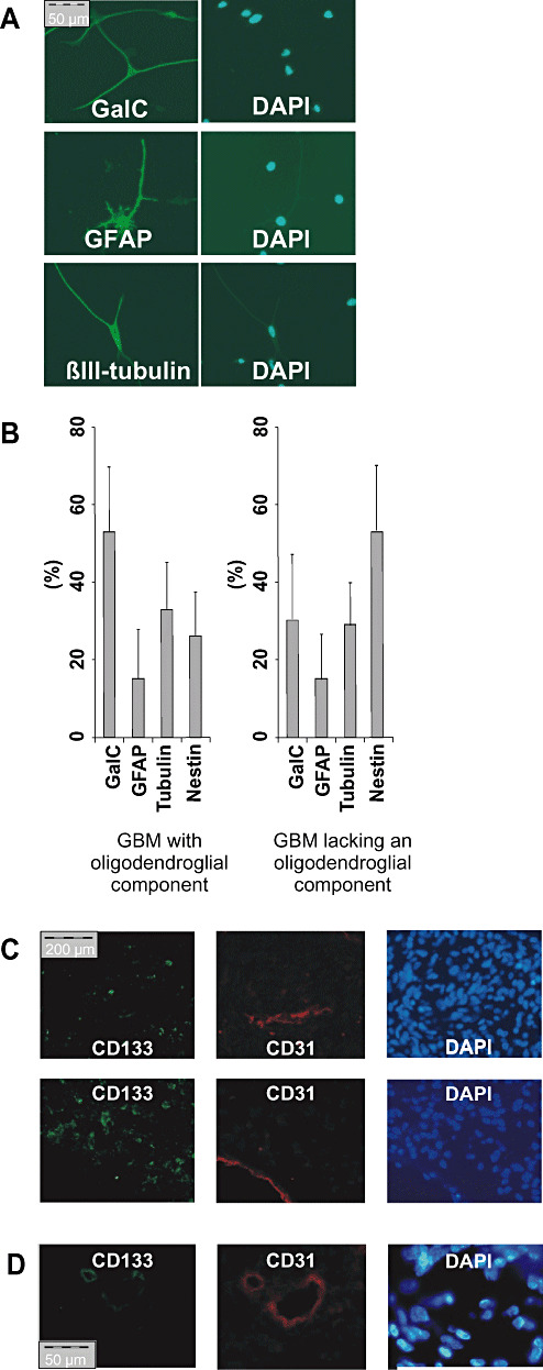
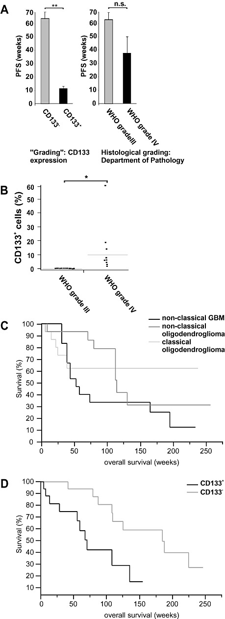
High-grade oligodendroglial tumors, that is, anaplastic oligodendroglial tumors and glioblastomas with oligodendroglial component, differ significantly in terms of prognosis and response to chemotherapy. Differentiation might be difficult because the histological differences are vague and reliable markers are not established. We correlated the presence of putative cancer stem cells (CSC) in high-grade oligodendroglial tumors (WHO grades III and IV) with clinical outcome. Tumors with favorable prognosis neither contained CSC nor did they show CD133 expression. Tumor cells resembled lineage-restricted progenitor cells with limited proliferative capacity and differentiation profile. In contrast, CD133 expression and stem cell-like tumor cells characterized tumors with poor prognosis. They showed neurosphere-like growth, differentiated into cells of all neural lineages, and were tumorigenic in nude mice. In our series, CSC and expression of CD133 predicted the clinical course of disease better than the histological grading. To confirm these results, we retrospectively analyzed 36 high-grade oligodendroglial tumors. Again, CD133 expression indicated shorter survival and predicted clinical outcome more reliable than the histological assessment. Our data show that detection of CSC and expression of CD133 is predictive of prognosis in high-grade oligodendroglial tumors. The presence or absence of CD133(+) CSC might explain the crucial biological difference between WHO grade III and IV oligodendroglial tumors.


Figures

Figure 1
Figure 1
Tumor cells of high‐grade oligodendroglial tumors with favorable prognosis resemble CD133 lineage‐restricted progenitor cells. A. The in vitro growth pattern of tumors with favorable prognosis was analyzed after 7 and 21 days. Representative photographs are shown. B. Tumor spheres in high‐grade oligodendroglial tumors with favorable prognosis did not grow infinitely. The relative average sphere size of representative tumors with favorable prognosis (R24, R42, gray bars) as compared with primary GBM lacking an oligodendroglial component (R28 and R44, black bars) is given. Sphere size was determined weekly (DIV = days in vitro). C. Cells within spheres differentiated almost exclusively into GalC (Galactocerebrosid C, oligodendroglial marker) and βIII‐tubulin positive (neuronal marker) but barely GFAP‐ (glial fibrillary acid protein, astrocytic marker) or nestin‐expressing cells (stem cell marker). A representative differentiation profile is shown.
Figure 2
Figure 2
Tumor cells of high‐grade oligodendroglial tumors with poor prognosis are characterized by CSC and CD133 expression. A. Tumors with poor prognosis showed non‐adherent neurosphere‐like growth with few spheres of continuously increasing size within multiple small spheres showing limited growth. The growth pattern of primary glioblastomas (GBM) lacking an oligodendroglial component (R44) is shown for comparison (lower row). Representative photographs at DIV 7 and 21 are shown. B. The relative average sphere size of tumors with poor prognosis (R40, R52, R30, gray bars) as compared with GBM lacking an oligodendroglial component (R28 and R44, black bars) is given. Sphere size was determined weekly. C. Cancer stem cell (CSC) lines derived from tumors with poor prognosis were sorted for CD133 expression and plated with 10 cells/well. The number of spheres formed by CD133+ and CD133 cells after 14 days is given (clonogenic index: spheres per 100 cells plated). D. GBMs with oligodendroglial component were driven by CSC reconstituting a new tumor when injected into immunodeficient mice (n = 4) and were similarly tumorigenic as CD133+ and CD133 CSC isolated from primary GBM lacking an oligodendroglial component (2).
Figure 3
Figure 3
CD133 + cells derived from high‐grade oligodendroglial tumors with poor prognosis differentiate into cells expressing all neural lineage markers. A. Spheres derived from single CD133+ tumor cells were stained as in Figure 1C for markers of all three neural lineages. B. A representative differentiation profile of CSC derived from a glioblastomas with (left) and without an oligodendroglial component (right) is given. C,D. Immunofluorescence staining against CD133 (green) and CD31 (red). (C) Two representative pictures of CD133+ tumors without CD133+/CD31+ cells. (D) One of 20 tumors investigated showed CD133+/CD31+ vessel‐like structures.
Figure 4
Figure 4
In high‐grade oligodendroglial tumors, CD133 expression indicates stem cell‐like tumor cells and is a negative prognostic marker. A. The progression‐free survival (PFS) of patients with CD133+ high‐grade oligodendroglial tumors was significantly shorter as compared to CD133 tumors (left panel, **P < 0.01, Wilcoxon rank test). The histological grading (Department of Pathology, University of Regensburg) did not significantly correlate with PFS (right panel, P = 0.74, Wilcoxon rank test). B. The CD133 expression in six anaplastic oligoastrocytomas (WHO grade III), four anaplastic oligodendrogliomas (WHO grade III), and 10 primary GBM lacking an oligodendroglial component (WHO grade IV) is given (*P < 0.05). C,D. Previously published microarrays and corresponding prognostic data were analyzed for CD133 expression (13). C. Tumors were classified by senior neuropathologists as classical anaplastic oligodendrogliomas, non‐classical anaplastic oligodendrogliomas and non‐classical GBM. A tumor was considered to be “classical” if it had a textbook‐like histological appearance [i.e. anaplastic oligodendroglioma or glioblastomas (GBM)] and if all neuropathologists involved agreed on the diagnosis. Otherwise, the tumor was classified as “non‐classical” oligodendroglioma or GBM. The histological diagnosis did not significantly correlate with survival (P > 0.10, Wilcoxon rank test). D. Tumors were grouped according to microarray data documenting the presence (i.e. “P call” in the array analysis) or absence (i.e. “A call” or “M call”) of CD133 mRNA expression. Patients with CD133 tumors survived significantly longer than patients with CD133+ tumors (P < 0.05, Wilcoxon rank test, n = 36).

Similar articles

Cited by

References

    1. Bao S, Wu Q, McLendon RE, Hao Y, Shi Q, Hjelmeland AB et al (2006) Glioma stem cells promote radioresistance by preferential activation of the DNA damage response. Nature 444:756–760. - PubMed
    1. Beier D, Hau P, Proescholdt M, Lohmeier A, Wischhusen J, Oefner PJ et al (2007) CD133+ and CD133‐ glioblastoma‐derived cancer stem cells show differential growth characteristics and molecular profiles. Cancer Res 67:4010–4015. - PubMed
    1. Bjerkvig R, Tysnes BB, Aboody KS, Najbauer J, Terzis AJ (2005) Opinion: the origin of the cancer stem cell: current controversies and new insights. Nat Rev Cancer 5:899–904. - PubMed
    1. Cairncross JG, Macdonald DR (1988) Successful chemotherapy for recurrent malignant oligodendroglioma. Ann Neurol 23:360–364. - PubMed
    1. Clarke MF (2005) Self‐renewal and solid‐tumor stem cells. Biol Blood Marrow Transpl 11:14–16. - PubMed