

Pineocytoma and pineal parenchymal tumors of intermediate differentiation presenting cytologic pleomorphism: a multicenter study
- PMID: 18371183
- PMCID: PMC8095592
- DOI: 10.1111/j.1750-3639.2008.00128.x
Pineocytoma and pineal parenchymal tumors of intermediate differentiation presenting cytologic pleomorphism: a multicenter study
Abstract
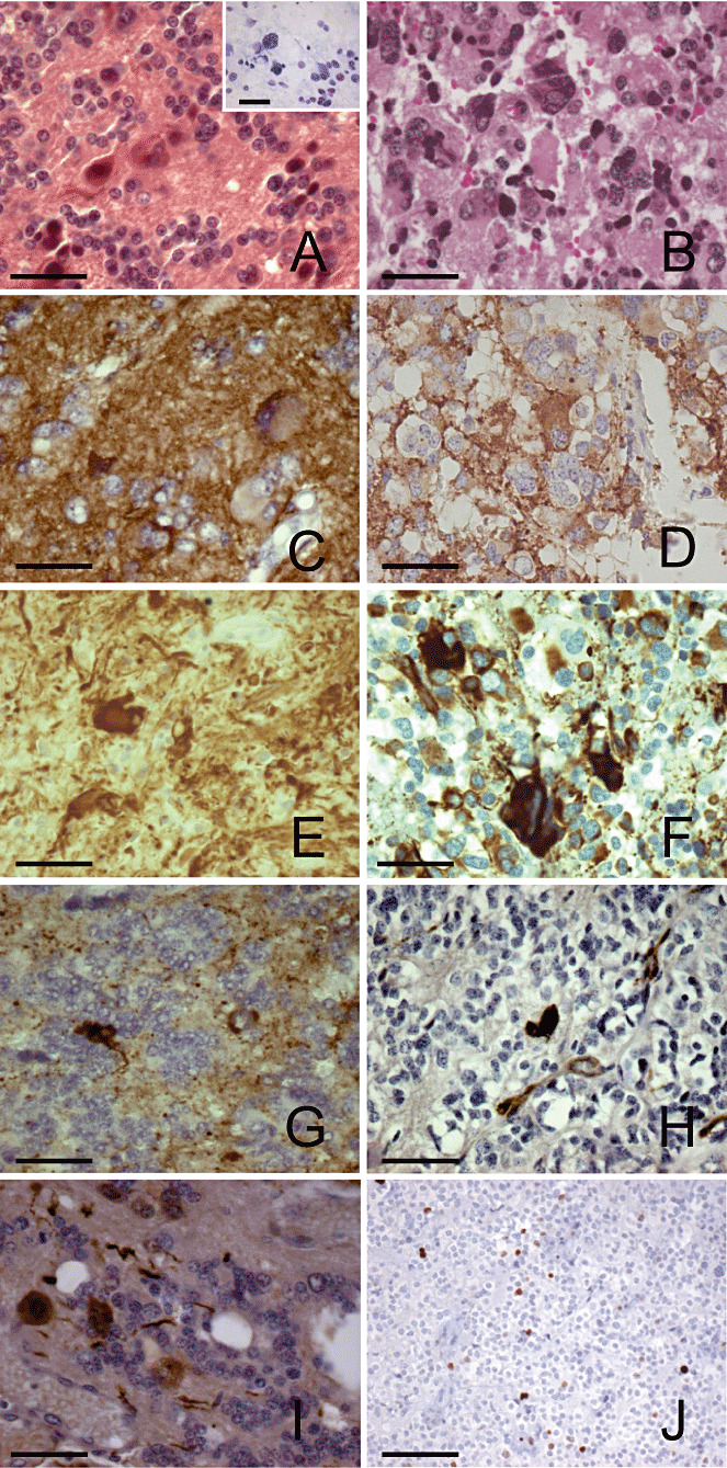
Cytologic pleomorphism has been described in a limited number of benign pineal tumors, namely pineocytoma (PC) and pineal parenchymal tumors (PPTs) of intermediate differentiation (PPTID). We examined the clinicopathologic features in a retrospective series of 14 cases (seven females and seven males aged from 10 to 65 years) of pleomorphic PPT. Seven cases were PC, with no mitoses and with areas of tumoral cells forming large pineocytomatous rosettes and other areas with giant cells containing hyperchromatic nuclei. The other seven were PPTID, presenting few mitoses (< or =2), a Ki67 proliferation index between 3% and 7%, and predominantly composed of small neoplastic cells and scattered giant cells, sometimes multinucleated. In the 14 tumors, the proportion of pleomorphic areas was variable. Most tumoral cells showed extensive neuronal differentiation with strong expression of neuron-specific enolase, synaptophysin and neurofilaments. Some of the neoplastic cells expressed S100 protein. The follow-up period ranged from 1.2 to 13 years and only one PC and one PPTID progressed after stereotactic biopsy or incomplete resection. The lack of invasiveness and the low proliferation index of these tumors suggest a benign clinical course despite the marked pleomorphism, the latter of which can lead to upgrading.
Figures
Similar articles
-
[Pineal Parenchymal Tumor with Marked Cytologic Pleomorphism: Is there a Correlation with the Malignancy Grade?].No Shinkei Geka. 2016 Jun;44(6):481-7. doi: 10.11477/mf.1436203314. No Shinkei Geka. 2016. PMID: 27270146 Japanese.
-
Clinicopathologic study of pineal parenchymal tumors of intermediate differentiation.World Neurosurg. 2014 May-Jun;81(5-6):783-9. doi: 10.1016/j.wneu.2013.02.007. Epub 2013 Feb 8. World Neurosurg. 2014. PMID: 23396072
-
Pineal parenchymal tumor of intermediate differentiation with cytologic pleomorphism.Neuropathology. 2006 Jun;26(3):212-7. doi: 10.1111/j.1440-1789.2006.00676.x. Neuropathology. 2006. PMID: 16771177
-
Pathology of pineal parenchymal tumors.Prog Neurol Surg. 2009;23:12-25. doi: 10.1159/000210050. Epub 2009 Mar 23. Prog Neurol Surg. 2009. PMID: 19329858 Review.
-
Pineal parenchymal tumors: a correlation of histological features with prognosis in 66 cases.Brain Pathol. 2000 Jan;10(1):49-60. doi: 10.1111/j.1750-3639.2000.tb00242.x. Brain Pathol. 2000. PMID: 10668895 Free PMC article. Review.
Cited by
-
Understanding and Managing Pineal Parenchymal Tumors of Intermediate Differentiation: An In-Depth Exploration from Pathology to Adjuvant Therapies.J Clin Med. 2024 Feb 23;13(5):1266. doi: 10.3390/jcm13051266. J Clin Med. 2024. PMID: 38592098 Free PMC article. Review.
-
Pineal parenchymal tumors of intermediate differentiation: in need of a stringent definition to avoid confusion. Scientific commentary on 'Genetical and epigenetical profiling identifies two subgroups of pineal parenchymal tumors of intermediate differentiation (PPTID) with distinct molecular, histological and clinical characteristics'.Acta Neuropathol. 2024 Feb 10;147(1):34. doi: 10.1007/s00401-024-02684-3. Acta Neuropathol. 2024. PMID: 38340187 Free PMC article. No abstract available.
-
Histopathology and molecular pathology of pediatric pineal parenchymal tumors.Childs Nerv Syst. 2023 Sep;39(9):2273-2284. doi: 10.1007/s00381-022-05637-x. Epub 2022 Aug 16. Childs Nerv Syst. 2023. PMID: 35972537 Review.
-
Pineal parenchymal tumor of intermediate differentiation: imaging spectrum of an unusual tumor in 11 cases.Neuroradiology. 2011 Aug;53(8):577-84. doi: 10.1007/s00234-010-0794-2. Epub 2010 Nov 16. Neuroradiology. 2011. PMID: 21080159
-
Binucleated and Multinucleated Neurons are Formed by Fusion.Bull Exp Biol Med. 2021 Aug;171(4):508-512. doi: 10.1007/s10517-021-05261-w. Epub 2021 Sep 20. Bull Exp Biol Med. 2021. PMID: 34542766
References
-
- Abe T, Inoue R, Isono M, Ishii K, Fujiki M, Kamida T et al (2006) Benign pleomorphic astrocytoma in the hypothalamus‐case report. Neurol Med Chir (Tokyo) 46:101–103. - PubMed
-
- Blumcke I, Wiestler OD (2002) Gangliogliomas: an intriguing tumor entity associated with focal epilepsies. J Neuropathol Exp Neurol 61:575–584. - PubMed
-
- Blumcke I, Giencke K, Wardelmann F, Beyenburg S, Kral T, Sarioglu N et al (1999) The CD34 epitope is expressed in neoplastic and malformative lesions associated with chronic, focal epilepsies. Acta Neuropathol 97:481–490. - PubMed
-
- Faillot T, Sichez JP, Capelle L, Kujas M, Fohanno D (1998) Ganglioglioma of the pineal region: case report and review of the literature. Surg Neurol 49:104–107. - PubMed
-
- Fèvre‐Montange M, Jouvet A, Privat K, Korf HW, Champier J, Reboul A et al (1998) Immunohistochemical, ultrastructural, biochemical and in vitro studies of a pineocytoma. Acta Neuropathol (Berl) 95:532–539. - PubMed
Publication types
MeSH terms
LinkOut - more resources
Full Text Sources
Medical
