

The receptor for granulocyte-colony stimulating factor (G-CSF) is expressed in radial glia during development of the nervous system
- PMID: 18371196
- PMCID: PMC2329616
- DOI: 10.1186/1471-213X-8-32
The receptor for granulocyte-colony stimulating factor (G-CSF) is expressed in radial glia during development of the nervous system
Abstract
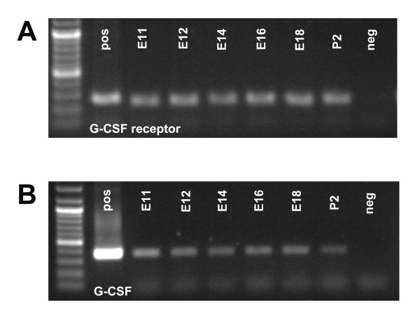
Background: Granulocyte colony-stimulating (G-CSF) factor is a well-known hematopoietic growth factor stimulating the proliferation and differentiation of myeloid progenitors. Recently, we uncovered that G-CSF acts also as a neuronal growth factor in the brain, which promotes adult neural precursor differentiation and enhances regeneration of the brain after insults. In adults, the receptor for G-CSF is predominantly expressed in neurons in many brain areas. We also described expression in neurogenic regions of the adult brain, such as the subventricular zone and the subgranular layer of the dentate gyrus. In addition, we found close co-localization of the G-CSF receptor and its ligand G-CSF. Here we have conducted a systematic expression analysis of G-CSF receptor and its ligand in the developing embryo.
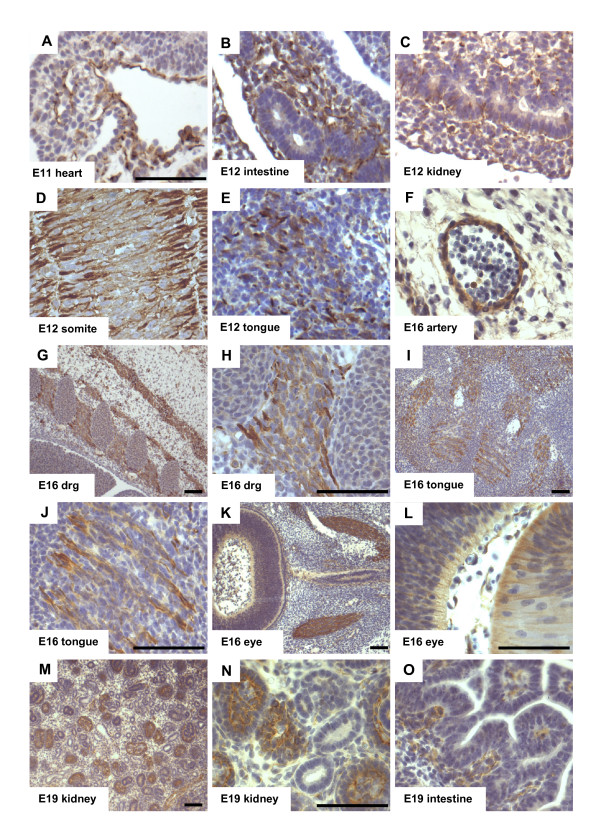
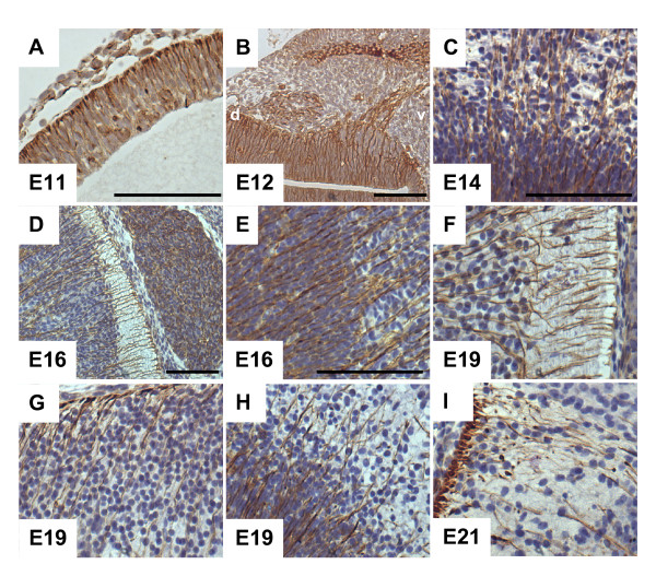
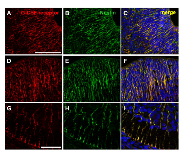
Results: Outside the central nervous system (CNS) we found G-CSF receptor expression in blood vessels, muscles and their respective precursors and neurons. The expression of the G-CSF receptor in the developing CNS was most prominent in radial glia cells.
Conclusion: Our data imply that in addition to the function of G-CSF and its receptor in adult neurogenesis, this system also has a role in embryonic neurogenesis and nervous system development.
Figures
Similar articles
-
Granulocyte-colony stimulating factor is neuroprotective in a model of Parkinson's disease.J Neurochem. 2006 May;97(3):675-86. doi: 10.1111/j.1471-4159.2006.03727.x. Epub 2006 Mar 29. J Neurochem. 2006. PMID: 16573658
-
Granulocyte-colony stimulating factor, granulocyte-macrophage colony stimulating factor, PIXY-321, stem cell factor, interleukin-3, and interleukin-7: receptor binding and effects on clonogenic proliferation in acute lymphoblastic leukemia.Leuk Lymphoma. 1994 Dec;16(1-2):79-88. doi: 10.3109/10428199409114143. Leuk Lymphoma. 1994. PMID: 7535143
-
Stem cell factor and granulocyte colony-stimulating factor promote neuronal lineage commitment of neural stem cells.Differentiation. 2012 Jan;83(1):17-25. doi: 10.1016/j.diff.2011.08.006. Epub 2011 Oct 5. Differentiation. 2012. PMID: 22099173
-
A role for G-CSF (granulocyte-colony stimulating factor) in the central nervous system.Cell Cycle. 2005 Dec;4(12):1753-7. doi: 10.4161/cc.4.12.2213. Epub 2005 Dec 27. Cell Cycle. 2005. PMID: 16258290 Review.
-
Regulation of myeloid development and function by colony stimulating factors.Dev Comp Immunol. 2004 May 3;28(5):509-54. doi: 10.1016/j.dci.2003.09.010. Dev Comp Immunol. 2004. PMID: 15062647 Review.
Cited by
-
Effects and safety of granulocyte colony-stimulating factor in healthy volunteers.Curr Opin Hematol. 2009 Jan;16(1):35-40. doi: 10.1097/MOH.0b013e328319913c. Curr Opin Hematol. 2009. PMID: 19057203 Free PMC article. Review.
-
Distribution of the hematopoietic growth factor G-CSF and its receptor in the adult human brain with specific reference to Alzheimer's disease.J Anat. 2014 Apr;224(4):377-91. doi: 10.1111/joa.12154. Epub 2014 Jan 5. J Anat. 2014. PMID: 24387791 Free PMC article.
-
Protein biomarkers of neural system.J Otol. 2019 Sep;14(3):77-88. doi: 10.1016/j.joto.2019.03.001. Epub 2019 Mar 23. J Otol. 2019. PMID: 31467504 Free PMC article. Review.
-
Interleukin-1 primes human mesenchymal stem cells towards an anti-inflammatory and pro-trophic phenotype in vitro.Stem Cell Res Ther. 2017 Apr 17;8(1):79. doi: 10.1186/s13287-017-0531-4. Stem Cell Res Ther. 2017. PMID: 28412968 Free PMC article.
-
Direct Potential Modulation of Neurogenic Differentiation Markers by Granulocyte-Colony Stimulating Factor (G-CSF) in the Rodent Brain.Pharmaceutics. 2022 Sep 2;14(9):1858. doi: 10.3390/pharmaceutics14091858. Pharmaceutics. 2022. PMID: 36145606 Free PMC article.
References
-
- Welte K, Platzer E, Gabrilove JL, Lu L, Levi E, Polivka A, Mertelsmann R, Moore MA. Purification to apparent homogeneity and biochemical characterization of human pluripotent hematopoietic colony-stimulating factor. Haematology and Blood Transfusion. 1985;29:398–401. - PubMed
-
- Schneider A, Kruger C, Steigleder T, Weber D, Pitzer C, Laage R, Aronowski J, Maurer MH, Gassler N, Mier W, Hasselblatt M, Kollmar R, Schwab S, Sommer C, Bach A, Kuhn HG, Schabitz WR. The hematopoietic factor G-CSF is a neuronal ligand that counteracts programmed cell death and drives neurogenesis. The Journal of clinical investigation. 2005;115:2083–2098. doi: 10.1172/JCI23559. - DOI - PMC - PubMed
MeSH terms
Substances
LinkOut - more resources
Full Text Sources
Molecular Biology Databases
