. 2008 Mar 27:6:13.
doi: 10.1186/1477-7827-6-13.

Angiogenic, hyperpermeability and vasodilator network in utero-placental units along pregnancy in the guinea-pig (Cavia porcellus)

Affiliations

Angiogenic, hyperpermeability and vasodilator network in utero-placental units along pregnancy in the guinea-pig (Cavia porcellus)

Gloria Valdés et al. Reprod Biol Endocrinol. .

Abstract

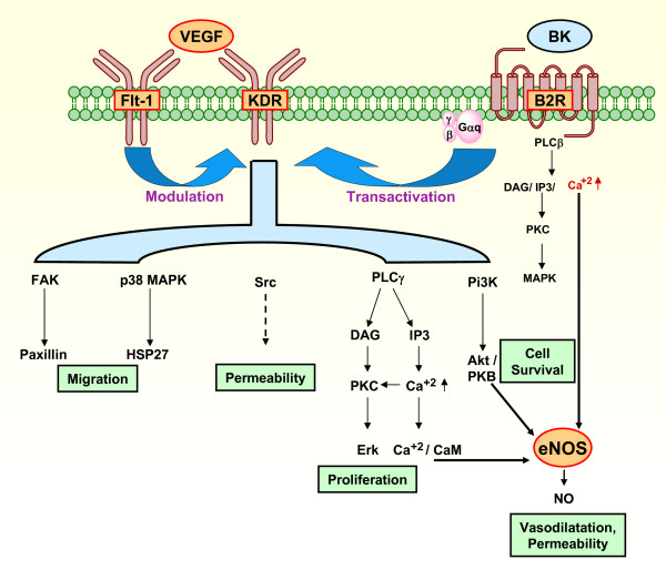
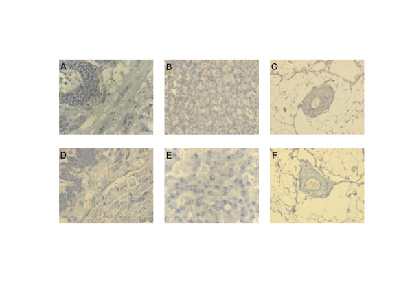
Background: The angiogenic and invasive properties of the cytotrophoblast are crucial to provide an adequate area for feto-maternal exchange. The present study aimed at identifying the localization of interrelated angiogenic, hyperpermeability and vasodilator factors in the feto-maternal interface in pregnant guinea-pigs.

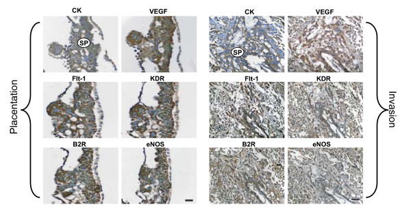
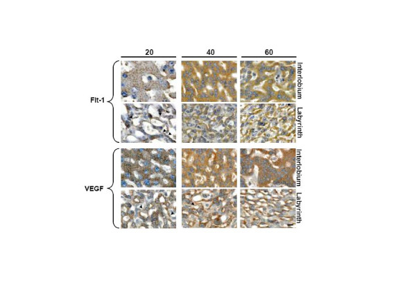
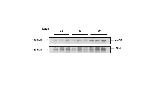
Methods: Utero-placental units were collected from early to term pregnancy. VEGF, Flt-1, KDR, B2R and eNOS were analyzed by immunohistochemistry, and the intensity of the signals in placenta and syncytial streamers was digitally analysed. Flt1 and eNOS content of placental homogenates was determined by western blotting. Statistical analysis used one-way analysis of variance and Tukey's Multiple Comparison post-hoc test.

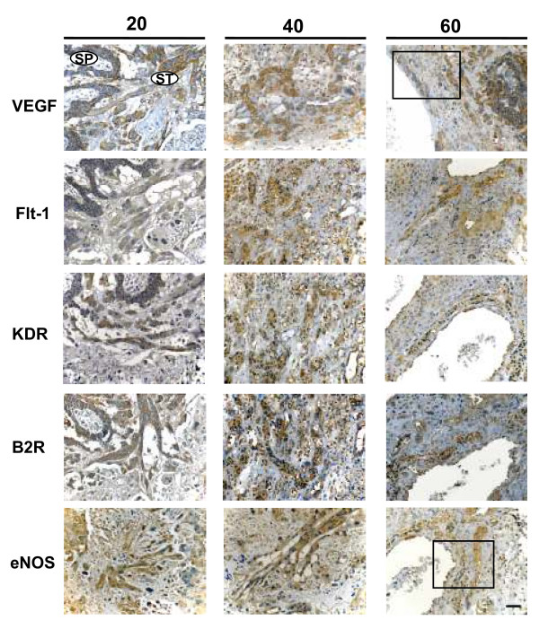
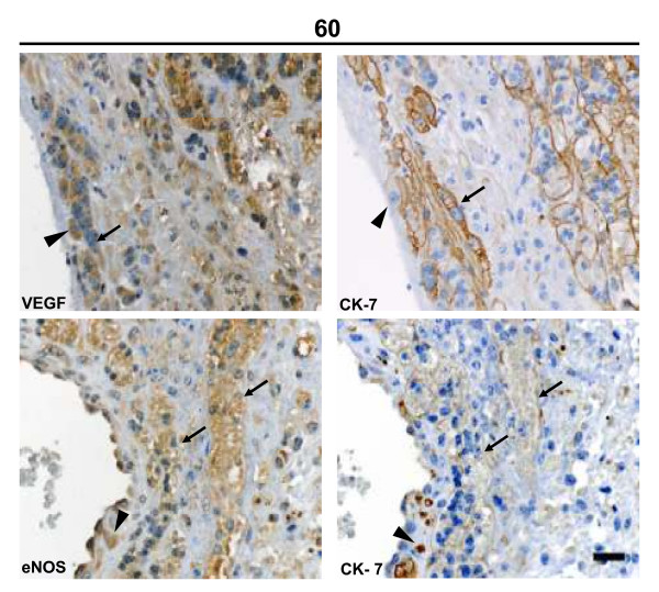
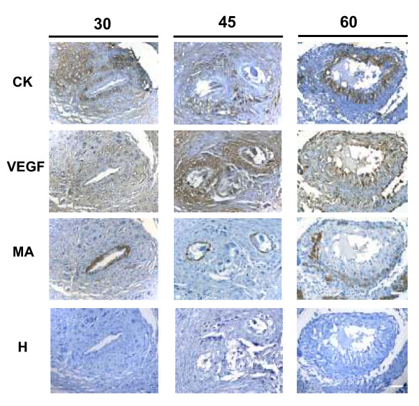
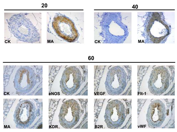
Results: In the subplacenta, placental interlobium and labyrinth VEGF, Flt-1, KDR, B2R and eNOS were expressed in all stages of pregnancy. Syncytial streamers in all stages of gestation, and cytotrophoblasts surrounding myometrial arteries in early and mid pregnancy - and replacing the smooth muscle at term - displayed immunoreactivity for VEGF, Flt-1, KDR, eNOS and B2R. In partly disrupted mesometrial arteries in late pregnancy cytotrophoblasts and endothelial cells expressed VEGF, Flt-1, KDR, B2R and eNOS. Sections incubated in absence of the first antibody, or in presence of rabbit IgG fraction and mouse IgG serum, yielded no staining. According to the digital analysis, Flt-1 increased in the placental interlobium in days 40 and 60 as compared to day 20 (P = 0.016), and in the labyrinth in day 60 as compared to days 20 and 40 (P = 0.026), while the signals for VEGF, KDR, B2R, and eNOS showed no variations along pregnancy. In syncytial streamers the intensity of VEGF immunoreactivity was increased in day 40 in comparison to day 20 (P = 0.027), while that of B2R decreased in days 40 and 60 as compared to day 20 (P = 0.011); VEGF, Flt-1, KDR, B2R and eNOS expression showed no variations. Western blots for eNOS and Flt-1 in placental homogenates showed no significant temporal differences along pregnancy.

Conclusion: The demonstration of different angiogenic, hyperpermeability and vasodilator factors in the same cellular protagonists of angiogenesis and invasion in the pregnant guinea-pig, supports the presence of a functional network, and strengthens the argument that this species provides an adequate model to understand human pregnancy.


Figures

Figure 1
Figure 1
Signaling pathways that contribute to VEGF induced angiogenesis, and a proposal for their participation in the development of the utero-placental interface by participating in proliferative, invasive, vasodilatory and permeability changes essential for cell invasion and angiogenesis. VEGF activates eNOS through pathways including Akt/PKB, Ca+2/CaM, and PKC [20-24]. Flt-1 may negatively regulate KDR, but might also promote its activity [20]. Bradykinin stimulates eNOS through Ca+2, induces EC to form tubes, and transactivates the KDR [26]. The factors studied have been depicted in orange areas surrounded by a red border, known mechanisms of downstream activation by interrupted arrows, and unknown mechanism of downstream activation by interrupted arrow. VEGF, vascular endothelial growth factor; BK, bradykinin; eNOS, endothelial nitric oxide synthase; PLC β, phospholipase C -β; PLCγ, phospholipase C -γ; DAG, diacylglycerol; IP3, inositol (1,4,5)-triphosphate; PKC, protein kinase C; MAPK, mitogen-activated protein kinase; FAK, focal adhesion kinase; PI3K, phosphoinositide 3-kinase; p38 MAPK, p38 mitogen-activated protein; Erk, extracellular regulated kinase; HSP27, heat protein; CaM, calmodulin; EC, endothelial cell.
Figure 2
Figure 2
In subsequent sections of a utero-placental unit obtained in day 15 of pregnancy, the multilayered subplacenta (SP) gave rise to the placental sprouts, and to the syncytial streamers that penetrate the adjacent decidua. The subplacenta, the placental sprouts and the syncytial streamers expressed VEGF, Flt-1, KDR, B2R and eNOS. Cytotrophoblasts were characterized by cytokeratin (CK) staining. Bar = 100 μm.
Figure 3
Figure 3
Syncytiotrophoblasts composing the interlobium and the labyrinth expressed Flt-1 and VEGF, in sections obtained in days 20, 40 and 60 of pregnancy at a high magnification (1000×). The immunoreactivity showed a granular pattern for interlobar Flt-1, and for interlobar and labyrinthine VEGF; labyrinthine Flt-1 displayed a diffuse cytoplasmic staining. Arrowheads highlight linear signal in endothelial cells. Bar = 100 μm.
Figure 4
Figure 4
Representative blot of placental homogenates from days 20, 40 and 60 of pregnancy for FLT-1 and eNOS. Purified human Flt-1 and eNOS yielded bands with approximate molecular weights of 180 and 140 kDa respectively. No significant differences were observed between the means of the 3 homogenates included in each study period, using one-way analysis of variance and Tukey's Multiple Comparison post-hoc test.
Figure 5
Figure 5
Syncytial streamers (ST) in the subplacenta (SP) and in the decidua expressed VEGF, Flt-1, KDR, B2R and eNOS in days 20, 40 and 60 of pregnancy. Bar = 100 μm. Rectangle defines the area shown at a higher magnification (400×) in Figure 6.
Figure 6
Figure 6
Syncytial streamers (arrows) in the vicinity of a decidual blood vessel in late pregnancy displayed granular VEGF and eNOS reactivity, and a linear pattern for cytokeratin-7 in the syncytial membrane. Intramural cytotrophoblasts (arrowheads) near the vessel lumen also expressed a granular eNOS reactivity, and intense intracytoplasmatic cytokeratin-7 staining. Bar = 100 μm.
Figure 7
Figure 7
Spiral arteries observed in sequential sections of myometrium obtained in days 30, 45 and 60 of pregnancy. Cytokeratin positive trophoblasts expressed VEGF. The remaining vascular smooth muscle layers were positive for muscle actin (MA), and have been almost replaced in day 60. Bar = 100 μm.
Figure 8
Figure 8
Mesometrial arteries obtained in days 20 and 40 of pregnancy showed in sequential sections an intact smooth muscle layer characterized by muscle actin (MA), and no cytokeratin positive cells. In day 60, intramural trophoblasts, characterized by cytokeratin (CK), expressed VEGF, Flt-1, KDR, B2R and eNOS, as did the swollen endothelial cells, identified by von Willebrand Factor (vWF). The remaining vascular smooth muscle, positive for muscle actin (MA), was disrupted. Bar = 100 μm.
Figure 9
Figure 9
Control sections at different days of pregnancy of the subplacenta and syncytial streamers (A, D), interlobium (B, E) and mesometrial arteries (C, F), which yielded no immunoreactivity incubated with non-specific IgG from mouse (1:50) and rabbit (1:100), species in which the antibodies were raised. Mouse IgG serum in (A, B, C) and rabbit IgG fraction in (D, E, F). Bar = 100 μm

Similar articles

Cited by

References

    1. Charnock-Jones DS, Kaufmann P, Mayhew TM. Aspects of human fetoplacental vasculogenesis and angiogenesis. I. Molecular regulation. Placenta. 2004;25:103–113. doi: 10.1016/j.placenta.2003.10.004. - DOI - PubMed
    1. Kaufmann P, Black S, Huppertz B. Endovascular trophoblast invasion: implications for the pathogenesis of intrauterine growth retardation and preeclampsia. Biol Reprod. 2003;69:1–7. doi: 10.1095/biolreprod.102.014977. - DOI - PubMed
    1. Kaufmann P, Mayhew TM, Charnock-Jones DS. Aspects of human fetoplacental vasculogenesis and angiogenesis. II. Changes during normal pregnancy. Placenta. 2004;25:114–126. doi: 10.1016/j.placenta.2003.10.009. - DOI - PubMed
    1. Kingdom J, Huppertz B, Seaward G, Kaufmann P. Development of the placental villous tree and its consequences for fetal growth. Eur J Obstet Gynecol Reprod Biol. 2000;92:35–43. doi: 10.1016/S0301-2115(00)00423-1. - DOI - PubMed
    1. Mayhew TM, Charnock-Jones DS, Kaufmann P. Aspects of human fetoplacental vasculogenesis and angiogenesis. III. Changes in complicated pregnancies. Placenta. 2004;25:127–139. doi: 10.1016/j.placenta.2003.10.010. - DOI - PubMed

Publication types

MeSH terms

Substances

LinkOut - more resources