. 2007 Jun 7;1(1):113-126.
doi: 10.1016/j.stem.2007.03.002.

Deletion of the developmentally essential gene ATR in adult mice leads to age-related phenotypes and stem cell loss

Affiliations

Deletion of the developmentally essential gene ATR in adult mice leads to age-related phenotypes and stem cell loss

Yaroslava Ruzankina et al. Cell Stem Cell. .

Abstract

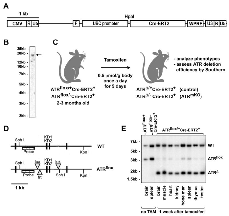
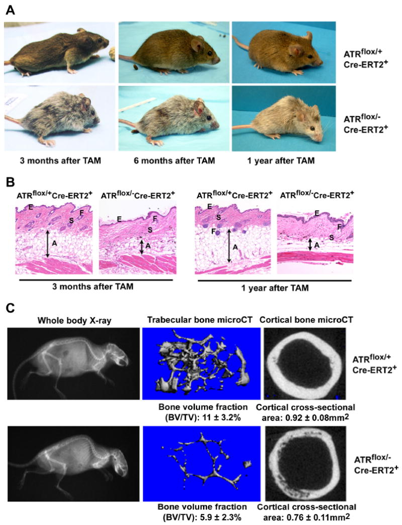
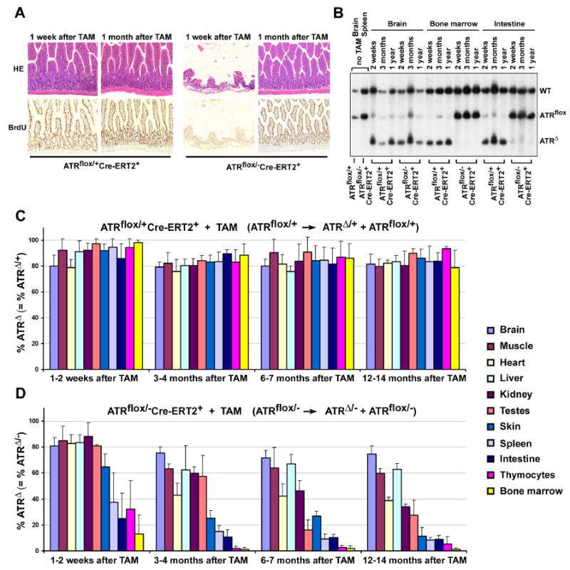
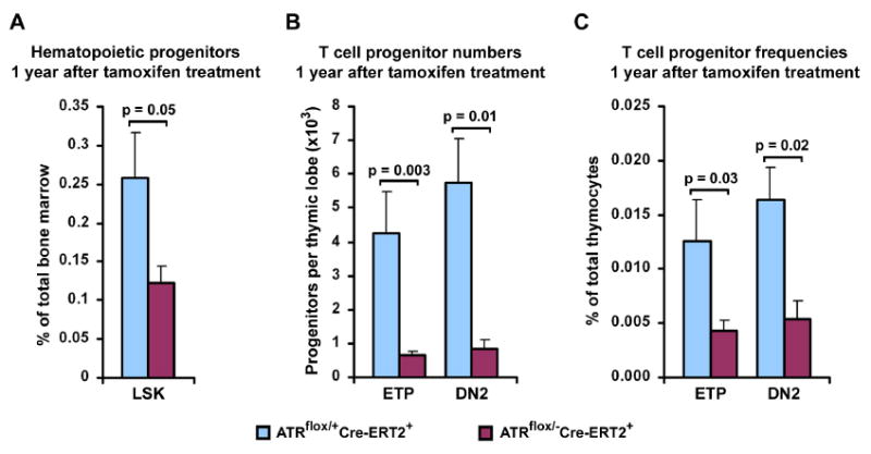
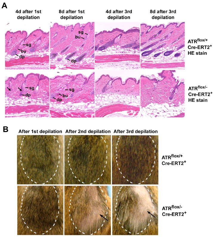
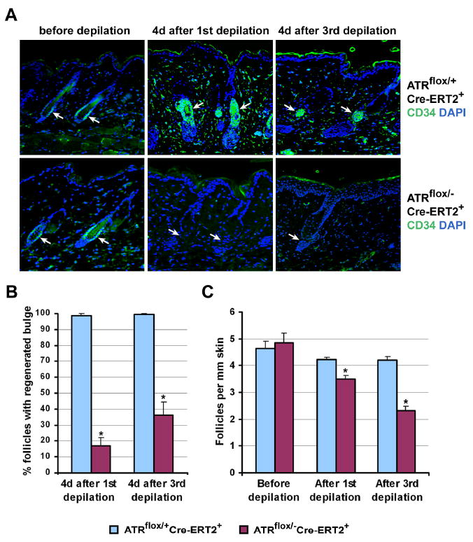
Developmental abnormalities, cancer, and premature aging each have been linked to defects in the DNA damage response (DDR). Mutations in the ATR checkpoint regulator cause developmental defects in mice (pregastrulation lethality) and humans (Seckel syndrome). Here we show that eliminating ATR in adult mice leads to defects in tissue homeostasis and the rapid appearance of age-related phenotypes, such as hair graying, alopecia, kyphosis, osteoporosis, thymic involution, fibrosis, and other abnormalities. Histological and genetic analyses indicate that ATR deletion causes acute cellular loss in tissues in which continuous cell proliferation is required for maintenance. Importantly, thymic involution, alopecia, and hair graying in ATR knockout mice were associated with dramatic reductions in tissue-specific stem and progenitor cells and exhaustion of tissue renewal and homeostatic capacity. In aggregate, these studies suggest that reduced regenerative capacity in adults via deletion of a developmentally essential DDR gene is sufficient to cause the premature appearance of age-related phenotypes.


Figures

Figure 1
Figure 1
A drug-inducible system to delete ATR in adult mice. (A) Lentiviral construct used to generate Cre-ERT2 fusion protein-expressing lentivirus. (B) A founder with a single copy integrant that expressed high levels of Cre-ERT2 was chosen to establish Cre-ERT2 lentitransgenic mouse line (arrow). (C) TAM treatment regimen used to stimulate recombination of the ATRflox allele. Mice were treated TAM at 2-3 months of age by oral gavage or intraperitoneal injection and analyzed subsequently at various time points. (D) Schematic of the ATRflox region (Brown and Baltimore, 2003). The kinase domain-encoding exons (KD1 & KD2) and probed region are shown. The null allele of ATR (ATR-) is wild-type in this probed region. (E) Southern blot of genomic DNA isolated from TAM-treated ATRflox/+Cre-ERT2+ mice. DNA samples from various tissues were digested with Sph I and Kpn I, Southern blotted and detected for the ATRflox region using the probe indicated in (D). Sph I ATRflox allele fragment = 3.1 kb, Sph I ATRΔ allele fragment = 1.8 kb.
Figure 2
Figure 2
ATR deletion leads to hair graying, alopecia, kyphosis and osteoporosis. (A) ATR deletion following TAM treatment leads to pervasive hair graying and patchy hair loss and kyphosis in ATRmKO mice. (B) Age-related abnormalities in the skin of ATRmKO mice. Thinning of the subcutaneous adipose layer (A), thickening of the epidermis (E), loss of hair follicles (F) and sebaceous gland cell hypertrophy (S) were observed in ATRmKO mice (n = 19) but not in control mice (n = 22). Sections from sex-matched mice are shown. (C) Increased kyphosis and osteoporosis in ATRmKO mice. Control and ATRmKO mice were X-rayed 1 year after TAM treatment (left panel). Increased kyphosis over controls was observed in all ATRmKO mice analyzed (n = 6). To analyze bone volume and cross-sectional area, femurs from ATRmKO and control mice were subjected to microCT analysis (4-7 mice analyzed/group). Trabecular bone in the distal metaphysic (middle panel) and cortical bone cross-sectional area (right panel) was imaged and analyzed. Bone volume/total volume (BV/TV) and cortical area are shown as mean ± SD. P ≤ 0.04 as calculated by Student's T-test.
Figure 3
Figure 3
Reduced thymopoiesis, accelerated thymic involution and failed spermatogenesis in ATRmKO mice. (A) Quantification of thymocytes at differing time points after ATR deletion. Thymi from ATRmKO and control animals (6-12 mice per group) were processed through mesh and thymocytes were counted by hemacytometer. Standard error bars are shown; P values were calculated by Student's T test. NS – not significant. (B) Premature thymic involution in ATRmKO mice. A significant reduction in the volume of stromal cortical layer, where early T cell precursors reside, was observed in all ATRmKO mice analyzed 1 year after ATR deletion (n = 3, double arrow). K5 – medullary epithelium, K8 – cortical epithelium. (C) Testicular degeneration in ATRmKO mice. Dramatic testicular atrophy was observed in all ATRmKO mice examined (n = 7), but not in control mice (n = 5). Pictures in (B) and (C) were taken at 100× magnification.
Figure 4
Figure 4
ATR deletion leads to loss of proliferating cells in mice. (A) Rapid loss and reconstitution of proliferating intestinal epithelial cells after ATR deletion. Mice were treated with BrdU in drinking water for up to 1 month following TAM treatment. Loss of proliferating intestinal epithelial cells was observed 1 week after ATR deletion in ATRmKO mice (n = 4), however, a full recovery was observed 3 weeks later (n = 5). Pictures were taken at 100× magnification. (B) ATRΔ/- cells are rapidly lost in proliferating tissues (bone marrow, intestine), but not in the brain of ATR knockout mice. Southern blot of genomic DNA isolated from ATRflox/+Cre-ERT2+ (control) and ATRflox/-Cre-ERT2+ (ATRmKO) mice treated or left untreated with TAM. Appearance of the ATRΔ allele represents ATRΔ/+ cells in control mice and ATRΔ/- cells in test mice. (C, D) Percentage of ATRΔ/+ cells remains constant (C), while ATRΔ/- cells are lost (D) in various tissues following lox recombination. Southern blot band intensities of lox recombined (ATRΔ) and unrecombined (ATRflox) were used to quantify the total percentage of cells that maintained a recombined copy of ATR (ATRΔ). Band intensities were quantified by phosphoimager, and mean percentages were calculated from the ratio ATRΔ over ATRΔ + ATRflox. For each tissue and time point, 2-8 mice were evaluated; each error bar indicates one standard deviation.
Figure 5
Figure 5
Decreased levels of hematopoietic and thymic progenitors in ATRmKO mice. (A) Quantification of hematopoietic LSK (Lin-Sca+c-Kithi) progenitors in the bone marrow of ATRflox/+Cre-ERT2+ (control) and ATRflox/-Cre-ERT2+ (ATRmKO) mice 1 year after TAM treatment. 5-9 mice per group were analyzed. (B, C) Quantification of T cell progenitors in the thymus of control and test mice. A decline in numbers (B) and frequencies (C) of ETPs (early T cell progenitors, Lin-CD25-c-Kithi) and DN2 (downstream double negative 2 progenitors, Lin-CD25+c-Kithi) was observed in the thymi of ATRmKO mice 1 year after TAM treatment (5-7 mice per group). Standard error bars are shown; P values were calculated by Mann-Whitney U test.
Figure 6
Figure 6
ATR deletion causes dysfunctional hair follicle regenerative cycling. (A) Formation of the new lower hair follicle was delayed after the 1st depilation (n = 5 mice for each time point) and frequently defective after the 3rd depilation in ATRmKO skin (n = 3 mice for each time point) (arrows). ATR was deleted in 50-55 day old mice by topical treatment of 4-OH-tamoxifen on 2-3 cm2 of dorsal skin. Four days later, hair shafts (telogen phase) were plucked in the treated areas. Second and third round of depilation were performed in subsequent telogen phases. H&E-stained histological sections of control and ATRmKO skins 4 and 8 days after depilation are shown, 4-10 sections were analyzed per mouse. Sg – sebaceous gland, bu – bulge, dp – dermal pappila. (B) Alopecia and graying of hair in ATRmKO skin 3-4 weeks after the 1st, 2nd and 3rd rounds of depilation (arrows). Depilated areas are marked by hatched lines. Defective hair regrowth was observed in all ATRmKO mice (n = 3-7 for each time point), but not in control mice (n = 3-8).
Figure 7
Figure 7
ATR deletion leads to bulge stem cell and follicle loss after depilation. (A) Bulge stem cells were detected by CD34 immunohistochemistry (green) in telogen phase (before depilation) and anagen phase (4 days after the 1st and 3rd depilations) follicles; nuclei were detected with DAPI (blue). Significant loss and delayed regeneration of follicle bulge stem cells were observed in ATRmKO skin following depilation, while the bulge regenerated normally in the control mice (arrows). All pictures were taken at 100× magnification. (B) Quantification of the data presented in (A) Two mice per group and 8 skin sections per mouse were used, and at least 12 follicles per section were analyzed. (C) Quantification of the average number of follicles per 1 mm of skin in control and ATRmKO mice before depilation and 4-6 weeks after each depilation. H&E stained skin sections were used. A significant decrease in follicle density in ATRmKO skin (n = 2-4 for each group) was observed. Error bars represent SEM. (*) p < 0.001 as calculated by Student's T-test.

Comment in

  • Stem cells and the rate of living.
    Krishnamurthy J, Sharpless NE. Krishnamurthy J, et al. Cell Stem Cell. 2007 Jun 7;1(1):9-11. doi: 10.1016/j.stem.2007.05.004. Cell Stem Cell. 2007. PMID: 18371325

Similar articles

Cited by

References

    1. Alonso L, Fuchs E. The hair cycle. J Cell Sci. 2006;119:391–393. - PubMed
    1. Arnold SR, Spicer D, Kouseff B, Lacson A, Gilbert-Barness E. Seckel-like syndrome in three siblings. Pediatr Dev Pathol. 1999;2:180–187. - PubMed
    1. Blanpain C, Lowry WE, Geoghegan A, Polak L, Fuchs E. Self-renewal, multipotency, and the existence of two cell populations within an epithelial stem cell niche. Cell. 2004;118:635–648. - PubMed
    1. Boscherini B, Iannaccone G, La Cauza C, Mancuso G, Girotti F, Finocchi G, Pasquino AM. Intrauterine growth retardation. A report of two cases with bird-headed appearance, skeletal changes and peripheral GH resistance. Eur J Pediatr. 1981;137:237–242. - PubMed
    1. Brown EJ, Baltimore D. ATR disruption leads to chromosomal fragmentation and early embryonic lethality. Genes Dev. 2000;14:397–402. - PMC - PubMed

Publication types

Substances