. 2008 Jan 10;2(1):83-9.
doi: 10.1016/j.stem.2007.10.007.

The p47 GTPase Lrg-47 (Irgm1) links host defense and hematopoietic stem cell proliferation

Affiliations

The p47 GTPase Lrg-47 (Irgm1) links host defense and hematopoietic stem cell proliferation

Carl G Feng et al. Cell Stem Cell. .

Abstract

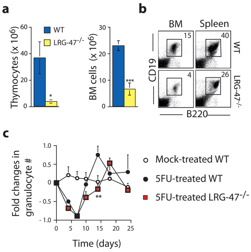
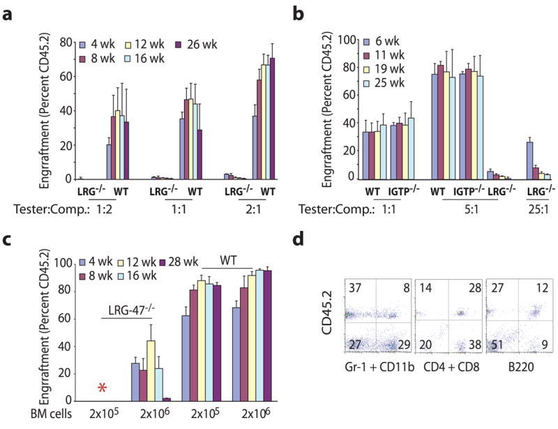
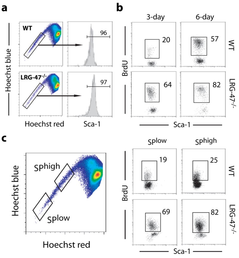
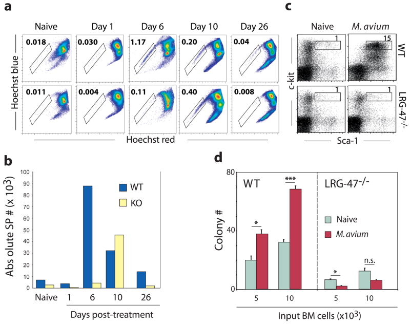
Hematopoietic stem cells (HSCs) are self-renewing bone marrow cells that give rise to all blood lineages and retain a remarkable capacity to proliferate in response to insult. Although some controls on HSC activation are known, little is understood about how this process is linked to natural signals. We report that the interferon-inducible GTPase Lrg-47 (Irgm1), previously shown to play a critical role in host defense, inhibits baseline HSC proliferation and is required for a normal HSC response to chemical and infectious stimuli. Overproliferating Lrg-47(-/-) HSCs are severely impaired in functional repopulation assays, and when challenged with hematopoietic ablation by 5-fluorouracil or infection with Mycobacterium avium, Lrg-47(-/-) mice fail to achieve the expected expansion response in stem and progenitor cell populations. Our results establish a link between the response to infection and HSC activation and demonstrate a novel function for a member of the p47 GTPase family.


Figures

Figure 1
Figure 1. Lrg-47 is required for hematopoietic recovery from insult
a–b. Following sublethal irradiation, Lrg-47 −/− mice show impaired recovery of hematopoietic organ cellularity, as well as reduced bone marrow and splenic B lymphocyte populations. Wild-type (WT; n=5) and Lrg-47 −/− (n=5) mice were subjected to sublethal irradiation, and were analyzed after 4 weeks for thymus and bone marrow cellularity (a) and percentage of spleen and bone marrow B cells (b; B220+CD19+ cells was identified by flow cytometric analysis). c. Recovery of peripheral blood elements following 5FU treatment is impaired, exemplified here by absolute granulocyte counts. The peripheral blood of mock-treated WT (n=5), 5FU-treated WT (n=9), and 5FU-treated Lrg-47 −/− (n=9) mice was analyzed periodically over a 24 day period; pooled data from two experiments with identical results is expressed as relative change from cohort baseline (absolute granulocyte counts were indexed to naïve values for each group). Differences between treated WT and KO are statistically significant at 14 days after injection, with p = 0.009.
Figure 2
Figure 2. Lrg-47 −/− HSCs show marked functional defects in bone marrow transplantation assays
a–b. Competitive bone marrow transplants. Whole bone marrow was isolated from CD45.2 WT or Lrg-47−/− donors (a–b) as well as IGTP−/− donors (b) and admixed in varying ratios with a constant number (2.5 × 10⁵) of whole bone marrow competitor cells from CD45.1 WT mice prior to injection into lethally irradiated recipient mice (CD45.1; n=7 recipients at the outset of the experiment). Engraftment was monitored by flow cytometric analysis of peripheral blood chimerism at the indicated timepoints. c+d. Non-competitive transplants. Whole bone marrow from CD45.2 WT or Lrg-47 −/− donors was transplanted without competitor into lethally irradiated CD45.1 recipients (n=5) at a dose of 2×10⁵ or 2×10⁶ cells, with peripheral blood chimerism monitored as before. (*) Recipients receiving 2×10⁵ KO cells died within two weeks of transplant. d. Representative FACS profile of Lrg-47 −/− engraftment, shown with myeloid and lymphoid lineage markers. Despite strikingly poor engraftment, analysis of Lrg-47 −/− HSC-derived peripheral blood at 16 weeks post-transplant demonstrates that these cells possess multilineage potential.
Figure 3
Figure 3. Lrg-47 negatively regulates HSC proliferation in the steady state
HSC proliferation status was assessed by in vivo BrdU labeling. Sca-1+ SP cells were purified from WT and Lrg-47 −/− mice after 3 or 6 days of BrdU exposure. The sorting strategy and percentage of Sca-1+ cells in the entire SP are illustrated in (a). After sorting, cells were permeabilized and stained for BrdU incorporation, prior to re-analysis by flow cytometry. Upon re-flow (b) cells were identified as Sca1+, and percentage of Sca-1+ SP cells that incorporated BrdU over the labeling period is indicated. In (c), WT and Lrg-47 −/− marrow was fractionated on the basis of SPlow (most primitive LT-HSC) vs. SPhigh gating, in addition to Sca-1 positivity, and analyzed as in (b). Data are representative of three independent experiments utilizing pools of bone marrow from 3–4 WT and Lrg-47 −/− mice in each.
Figure 4
Figure 4. Lrg-47 −/− HSCs are impaired in their response to both chemotherapeutic and microbial stress
a. The frequency of HSCs (SP cells) in wild-type and Lrg-47 −/− mice was determined in both naïve animals and mice that had been treated with 5FU 1, 6, 10, or 26 days prior to analysis. The frequency of SP cells (gate) is indicated as a percentage of live cells. b. By multiplying the observed frequency of HSCs by the overall bone marrow cellularity, it is possible to discern changes in the absolute number of HSCs following 5FU treatment, reported here as absolute SP#/hindlimb; following 5FU treatment, KO mice exhibit severe ablation of the HSC compartment. Data in a–b are representative of multiple experiments, and were obtained from marrow pools of 2–3 mice per data point. c. Wild-type and knockout animals were infected intravenously with M. avium, and sacrificed at 4 weeks post-infection. The percentage of the KSL population is indicated on Lin- gated plots. d. 4 weeks after infection, whole bone marrow from wild-type and knockout animals was plated in methylcellulose, and colony formation was assessed 12 days later. Infection induced a statistically significant increase in progenitor activity in wild-type marrow (p=0.0125 at 5k; p=0.0003 at 10k). Infection actually induced a drop in knockout colony-forming activity (p=0.0231 at 5k; p=0.0940 at 10k). Data is representative of two experiments with identical results.

Comment in

Similar articles

Cited by

References

    1. Genome-wide association study of 14,000 cases of seven common diseases and 3,000 shared controls. Nature. 2007;447:661–678. Wellcome Trust Case Control Consortium. - PMC - PubMed
    1. Butcher BA, Greene RI, Henry SC, Annecharico KL, Weinberg JB, Denkers EY, Sher A, Taylor GA. p47 GTPases regulate Toxoplasma gondii survival in activated macrophages. Infect Immun. 2005;73:3278–3286. - PMC - PubMed
    1. Camargo FD, Chambers SM, Drew E, McNagny KM, Goodell MA. Hematopoietic stem cells do not engraft with absolute efficiencies. Blood. 2006;107:501–507. - PMC - PubMed
    1. Collazo CM, Yap GS, Sempowski GD, Lusby KC, Tessarollo L, Woude GF, Sher A, Taylor GA. Inactivation of Lrg-47 and IRG-47 reveals a family of interferon gamma-inducible genes with essential, pathogen-specific roles in resistance to infection. J Exp Med. 2001;194:181–188. - PMC - PubMed
    1. Dufour C, Corcione A, Svahn J, Haupt R, Battilana N, Pistoia V. Interferon gamma and tumour necrosis factor alpha are overexpressed in bone marrow T lymphocytes from paediatric patients with aplastic anaemia. Br J Haematol. 2001;115:1023–1031. - PubMed

Publication types

MeSH terms