. 2008 Feb 7;2(2):183-9.
doi: 10.1016/j.stem.2007.11.002.

Transparent adult zebrafish as a tool for in vivo transplantation analysis

Affiliations

Transparent adult zebrafish as a tool for in vivo transplantation analysis

Richard Mark White et al. Cell Stem Cell. .

Abstract

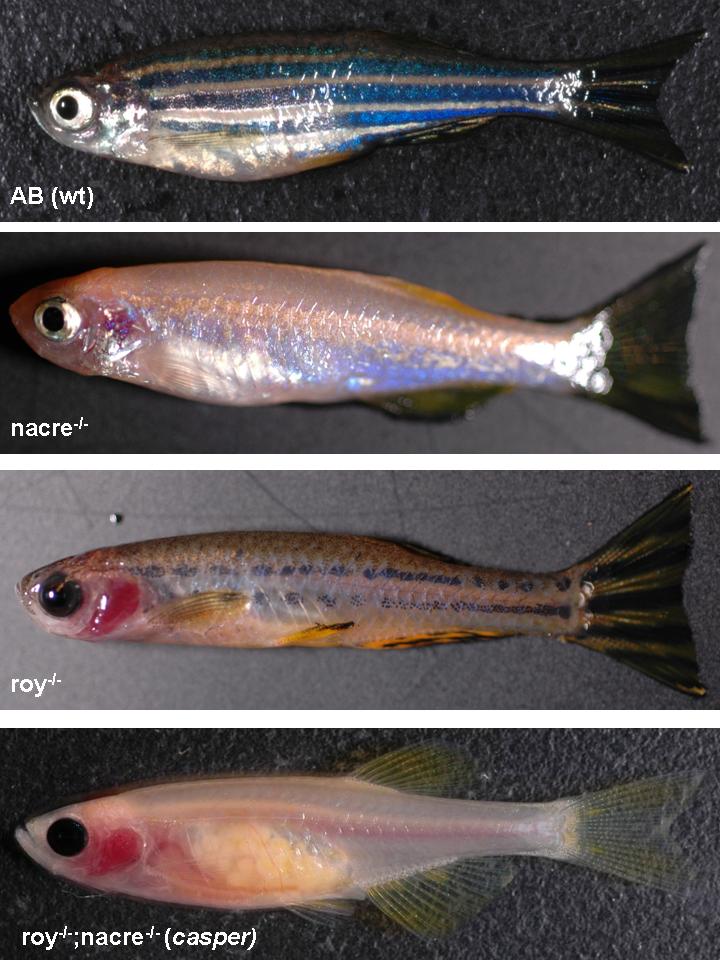
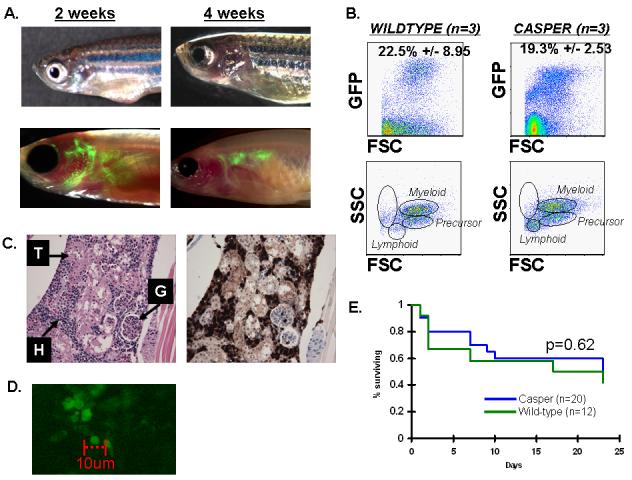
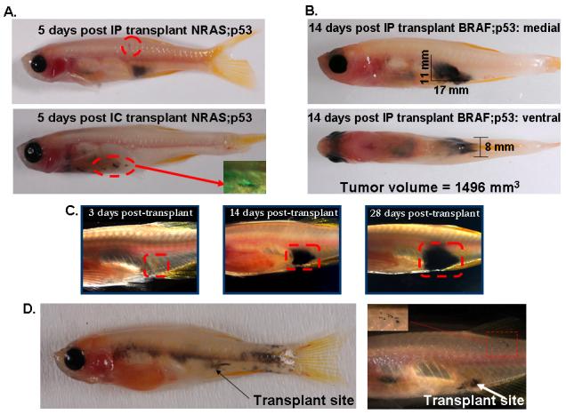
The zebrafish is a useful model for understanding normal and cancer stem cells, but analysis has been limited to embryogenesis due to the opacity of the adult fish. To address this, we have created a transparent adult zebrafish in which we transplanted either hematopoietic stem/progenitor cells or tumor cells. In a hematopoiesis radiation recovery assay, transplantation of GFP-labeled marrow cells allowed for striking in vivo visual assessment of engraftment from 2 hr-5 weeks posttransplant. Using FACS analysis, both transparent and wild-type fish had equal engraftment, but this could only be visualized in the transparent recipient. In a tumor engraftment model, transplantation of RAS-melanoma cells allowed for visualization of tumor engraftment, proliferation, and distant metastases in as little as 5 days, which is not seen in wild-type recipients until 3 to 4 weeks. This transparent adult zebrafish serves as the ideal combination of both sensitivity and resolution for in vivo stem cell analyses.


Figures

Figure 1
Figure 1
Zebrafish pigmentation mutants exhibit defects in specific neural crest derived populations. A wild-type zebrafish (1A) demonstrates alternating patterns of deeply pigmented stripes comprised largely of melanocytes. The interstripe regions, devoid of melanocytes, contain primarily reflective iridophores and yellowish xanthophores. The nacre mutant (1B), due to loss of mitf, shows a loss of embryonic and adult melanocytes, with a compensatory increase in iridophore numbers. The roy orbison mutant (1C), whose genetic defect is currently undefined, shows a striking abnormality of eye pigmentation, a severe disruption of melanocyte numbers/patterning, and a complete loss of the iridophore layer. The compound roy;nacre double homozygous mutant (1D), which we have named casper, shows the effect of combined melanocyte and iridophore loss, in which the body of the adult fish is largely transparent due to loss of light absorption and reflection.
Figure 2
Figure 2
Transverse section of wild-type (2A) and casper (roy;nacre) eyes (2B) demonstrates that the uniformly pigmented eye in the mutant line is primarily due to a loss of reflective iridophores in the sclera (arrow shows iridophores in wild-type fish), which then exposes the black underlying pigmented retinal epithelium. The remaining eye structures appear intact. Transverse sectioning of the adult skin from wild-type (2C) and casper (2D) shows a thick layer of iridophore crystals in the hypodermis of the wild-type adult (arrow) but a complete loss of this cell layer in the casper skin. Light that would normally be intercepted by epidermal/dermal melanocytes can penetrate deeply into the hypodermis of the mutant, and is not reflected away due to the lack of iridophore crystals. This allows for deep tissue penetration of normal wavelength length. M=muscle layer; D=dermis; Ir=iridophores, C=corneal surface.
Figure 3
Figure 3
Whole kidney marrow from beta actin:GFP labeled donors was transplanted into either wild-type (n=6) or casper (n=6) irradiated recipients via intra-cardiac (IC) injection of 100,000 cells. Fish were imaged from 4 hours until 5 weeks post transplant at once weekly intervals. At 2 weeks (3A, left), GFP positive cells can be seen to circulate and home to the region near the gills and head kidney of the recipient only in the casper line. By 4 weeks (3A, right), a population of GFP positive cells is tightly localized to the zebrafish kidney, where most adult hematopoietic tissue is known to reside. The GFP positive cells can only been seen in the transparent casper recipient. To confirm that the GFP labeled cells represented true hematopoietic cells, whole kidney was isolated from wild-type and casper recipients at 4 weeks, and subject to FACS analysis (3B). Both the wild-type (3B, left) and transparent mutant (3B, right) repopulated their kidney marrow with a full repertoire of hematopoietic lineages. In both types of recipients, a subset of the sorted cells were GFP positive (19-22%), as is expected in a mosaic transplantation assay. In (3C), histological analysis of the casper transplant recipient is shown at 4 weeks post transplant. H&E staining (3C, left) shows a robust population of hematopoietic cells (labeled “H”) intertwined with normal kidney glomeruli (labeled “G”) and tubules (labeled “T”). Immunohistochemistry using an anti-GFP antibody (3C, right) demonstrates that only the hematopoietic cells are strongly GFP positive (brown staining) whereas the kidney tubules and glomeruli are negative. Confocal laser scanning was used to examine a transplant recipient at 4 weeks post transplant (4D), and demonstrates easily discernible single GFP positive cells in the kidney marrow space. Survival after transplantation was identical in wild-type versus casper mutants (4E).
Figure 4
Figure 4
mitf-NRAS-GFP;p53-/-(4A) driven melanoma cells were transplanted into casper adults into either the peritoneum (IP, n=6) or via intracardiac injection (IC, n=5) at a dose of 200,000 cells. By 5 days post IP transplant of NRAS-GFP cells (4A, top), a large deeply pigmented intra-abdominal mass could be seen (n=3/6 fish) as well as spread to the dorsal epidermal scales (circled area). After IC injection of NRAS-GFP cells (4A, bottom), the cells proliferated and enlarged along the needle tract but did not appear to spread to distant sites. The inset, taken at 10 days post-transplant, shows that the cells continue to be strongly GFP positive. In Figure 4B, IP transplantation of mitf-BRAF;p53-/- melanoma cells demonstrates 14 day engraftment within the peritoneal cavity. Mediolateral and ventral views allow for calculation of 3-dimensional tumor volume. In 4C, repeated imaging of the same fish over a 1 month period demonstrates gradual increase in tumor volume without the need for sacrifice of the recipient. In a study of 24 transplant recipient fish, 37.5% were found to develop distant metastasis, examples of which are shown in 4D. Single migratory (stellate appearing) melanoma cells (4D, right, inset box) have migrated far from the transplantation site and embedded in the dorsal skin.

References

    1. Al-Hajj M, Wicha MS, Benito-Hernandez A, Morrison SJ, Clarke MF. Prospective identification of tumorigenic breast cancer cells. Proceedings of the National Academy of Sciences of the United States of America. 2003;100:3983–8. - PMC - PubMed
    1. Beckmann N, Kneuer R, Gremlich HU, Karmouty-Quintana H, Ble FX, Muller M. In vivo mouse imaging and spectroscopy in drug discovery. NMR Biomed. 2007;20:154–185. - PubMed
    1. Burns CE, Traver D, Mayhall E, Shepard JL, Zon LI. Hematopoietic stem cell fate is established by the Notch-Runx pathway. Genes & Development. 2005;19:2331–42. - PMC - PubMed
    1. Cao F, Lin S, Xie X, Ray P, Patel M, Zhang X, Drukker M, Dylla SJ, Connolly AJ, Chen X, et al. In vivo visualization of embryonic stem cell survival, proliferation, and migration after cardiac delivery. Circulation. 2006;113:1005–1014. - PMC - PubMed
    1. Clothier J, Lythgoe JN. Light-induced colour changes by the iridophores of the Neon tetra, Paracheirodon innesi. J. Cell. Sci. 1987;88(Pt 5):663–668. - PubMed

Publication types

LinkOut - more resources