. 2008 Jun 6;283(23):16084-92.
doi: 10.1074/jbc.M710223200. Epub 2008 Mar 27.

CUL2 is required for the activity of hypoxia-inducible factor and vasculogenesis

Affiliations

CUL2 is required for the activity of hypoxia-inducible factor and vasculogenesis

Yutaka Maeda et al. J Biol Chem. .

Abstract

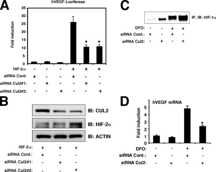
CULLIN 2 (CUL2) is a component of the ElonginB/C-CUL2-RBX-1-Von Hippel-Lindau (VHL) tumor suppressor complex that ubiquitinates and degrades hypoxia-inducible factor alpha (HIFalpha). HIFalpha is a transcription factor that mediates the expression of hypoxia-sensitive genes, including vascular endothelial growth factor (VEGF), which in turn regulates vasculogenesis. Whereas CUL2 participates in the degradation of HIFalpha, the potential role of CUL2 in the regulation of other cellular processes is less well established. In the present study, suppression of CUL2 expression by Cul2 siRNA inhibited HIFalpha transcriptional activation of the VEGF gene in vitro, indicating that CUL2 plays a role distinct from its known function in HIFalpha degradation. Because ARNT heterodimerizes with HIFalpha, we assessed whether CUL2 influenced ARNT expression. Cul2 siRNA inhibited the expression of endogenous ARNT. Ectopically expressed ARNT reversed the inhibition of HIF activity by Cul2 siRNA in the VEGF promoter, suggesting that CUL2 regulates HIF activation through ARNT. In 786-O cells lacking VHL, Cul2 siRNA suppressed the expression of both ARNT and VEGF, indicating that CUL2 regulates HIF activity independently of VHL. In transgenic zebrafish expressing GFP driven by the Flk promoter (a known HIF target), zCul2 morpholino blocked embryonic vasculogenesis in a manner similar to that caused by inhibition of VEGF-A. In the zebrafish embryos, zCul2 inhibited the expression of CUL2, VEGF, and Flk-GFP protein, indicating that CUL2 is required for expression of other vasculogenic HIF targets. Taken together, CUL2 is required for normal vasculogenesis, at least in part mediated by its regulation of HIF-mediated transcription.


Figures

FIGURE 1.
FIGURE 1.
CUL2 is required for HIFα-dependent activation of the VEGF promoter. A, CUL2 siRNA inhibited HIF-2α activity. H441 cells were transfected with 1 μg of hVEGF luciferase construct (hVEGF-Luciferase), 1 μg of pCMV.β-galactosidase, 1 μg of HIF-2α expression vector (pcDNA3.1.HIF-2α), 100 pmol of siRNA negative control (siRNA Cont.), or Cul2-targeting siRNA (siRNA Cul2) as indicated. The cells were incubated for 48 h before harvest. Results are presented as fold activation of light units normalized to β-galactosidase activity relative to control constructs. B, Cul2 siRNA effectively suppressed CUL2 protein expression. Cell lysates prepared in A were blotted with anti-CUL2 and anti-HIF-2α to identify CUL2 and HIF-2α. Anti-β-actin was used as a negative control. C, Cul2 siRNA stabilized the HIF-1α protein. HeLa cells were transfected with 100 pmol of siRNA negative control (siRNA Cont.) or Cul2-targeting siRNA (siRNA Cul2#1) as indicated. 24 h after transfection, 300 μm DFO was added to the transfected cells and incubated for 30 h before cell harvest. The immunoprecipitates obtained with anti-HIF-1α antibody (IP) were separated by SDS-PAGE, transferred to the membrane, and blotted with anti-HIF-1α (IB) as described under “Experimental Procedures.” D, Cul2 siRNA inhibited DFO-mediated VEGF induction. HeLa cells were transfected with 100 pmol of siRNA negative control (siRNA Cont.) or Cul2-targeting siRNA (siRNA Cul2#1) as indicated. 24 h after transfection, 300 μm DFO was added, and the cells were incubated for 30 h before cell harvest. Results of real-time PCR are presented as fold activation of VEGF mRNA normalized to 18 S rRNA.
FIGURE 2.
FIGURE 2.
RBX1-CUL2 is required for HIF-2α-mediated activation of the VEGF promoter. A, Rbx1 siRNA suppressed RBX1 and CUL2 protein expression. HeLa cells were transfected with 100 pmol of siRNA negative control (siRNA Cont.), Cul2-targeting siRNA (siRNA Cul2#1), or Rbx1-targeting siRNA (siRNA Rbx1). Cell lysates were blotted with anti-RBX1 and anti-CUL2 to identify RBX1 and CUL2. Anti-β-actin was used as a negative control. B, Rbx1 siRNA suppressed HIF-2α activity. H441 cells were transfected with 1 μg of the hVEGF luciferase construct (hVEGF-Luciferase), 1 μg of pCMV.β-galactosidase, 1 μg of HIF-2α expression vector (pcDNA3.1.HIF-2α), 100 pmol of siRNA negative control (siRNA Cont.), or Rbx1-targeting siRNA (siRNA Rbx1) as indicated. Results are presented as fold activation of light units normalized to β-galactosidase activity relative to control constructs. C, HIF-2α bound to TIP49 but not TIP60. Immunoprecipitates (IP) were prepared from MLE-15 (mouse lung epithelial cells) after transfection with FLAG-HIF-2α or FLAG empty control vector. The immunoprecipitates obtained with anti-FLAG antibody (IP: FLAG) were separated by SDS-PAGE, transferred to the membrane, and blotted with anti-FLAG, anti-TIP49, and anti-TIP60 as described under “Experimental Procedures.” D, both Cul2 siRNA and Tip49 siRNA were target-specific. HeLa cells were transfected with 100 pmol of siRNA negative control (siRNA Cont.), Cul2-targeting siRNA (siRNA Cul2#1), or Tip49-targeting siRNA (siRNA Tip49). Cell lysates were blotted with anti-CUL2 and anti-TIP49 to identify CUL2 and TIP49. *, nonspecific band. Arrow, TIP49-specific band. E, Tip49 did not influence DFO-mediated VEGF transcription. HeLa cells were transfected with 1μg of the hVEGF luciferase construct (hVEGF-Luciferase), 1μg of pCMV.β-galactosidase, 100 pmol of siRNA negative control (siRNA Cont.), Cul2-targeting siRNA (siRNA Cul2#1), or Tip49-targeting siRNA (siRNA Tip49) as indicated. 24 h after transfection, 300 μm DFO was added to the transfected cells and incubated for 30 h before cell harvest. Results are presented as fold activation of light units normalized to β-galactosidase activity relative to control constructs.
FIGURE 3.
FIGURE 3.
CUL2 influences HIF-2α activity through ARNT. A, ARNT siRNA suppressed ARNT protein expression. H441 cells were transfected with 100 pmol of siRNA negative control (siRNA Cont.) or ARNT-targeting siRNA (siRNA ARNT). Cell lysates were blotted with anti-ARNT and anti-CUL2 to identify ARNT and CUL2. Anti-β-actin was used as a negative control. B, ARNT siRNA inhibited HIF-2α activity. H441 cells were transfected with 1 μg of the hVEGF luciferase construct (hVEGF-Luciferase), 1 μg of pCMV.β-galactosidase, 1 μg of HIF-2α expression vector (pcDNA3.1.HIF-2α), 100 pmol of siRNA negative control (siRNA Cont.), or ARNT-targeting siRNA (siRNA ARNT) as indicated. Results are presented as fold activation of light units normalized to β-galactosidase activity relative to control constructs. C, Cul2 siRNA suppressed CUL2 and ARNT protein expression. HeLa cells were transfected with 100 pmol of siRNA negative control (siRNA Cont.) or Cul2-targeting siRNA (siRNA Cul2#1). Cell lysates were blotted with anti-CUL2 and anti-ARNT to identify CUL2 and ARNT. Anti-β-actin was used as a negative control. D, Cul2 siRNA suppressed ARNT mRNA expression. HeLa cells were transfected with 100 pmol of siRNA negative control (siRNA Cont.) or Cul2-targeting siRNA (siRNA Cul2#1) as indicated. The cells were incubated for 48 h before cell harvest. Results of real-time PCR are presented as fold activation of ARNT mRNA normalized to 18 S rRNA. E, ectopically expressed ARNT reversed the effect of Cul2 siRNA on HIF-2α activity. H441 cells were transfected with 1 μg of the hVEGF luciferase construct (hVEGF-Luciferase), 1 μg of pCMV.β-galactosidase, 1 μg of HIF-2α expression vector (pcDNA3.1.HIF-2α), 1 μg of ARNT expression vector (pcDNA3.1ARNT), 100 pmol of siRNA negative control (siRNA Cont.), or Cul2-targeting siRNA (siRNA Cul2#1) as indicated. Results are presented as fold activation of light units normalized to β-galactosidase activity relative to control constructs.
FIGURE 4.
FIGURE 4.
CUL2 is required for ARNT and VEGF mRNA expression in 786-O (VHL defective) cells. A, Cul2 siRNA suppressed CUL2, ARNT and HIF-2α protein expression. 786-O cells were transfected with 100 pmol of siRNA negative control (siRNA Cont.) or Cul2-targeting siRNA (siRNA Cul2#1). Cell lysates were blotted with anti-CUL2, anti-ARNT, and anti-HIF-2α to identify CUL2, ARNT, and HIF-2α. Anti-β-actin and anti-GAPDH were used as negative controls. B, Cul2 siRNA suppressed ARNT and VEGF mRNA expression. 786-O cells were transfected with 100 pmol of siRNA negative control (siRNA Cont.), Cul2-targeting siRNA (siRNA Cul2#1), or ARNT-targeting siRNA (siRNA ARNT) as indicated. The cells were incubated for 72 h before cell harvest. Results of real-time PCR are presented as fold activation of ARNT and VEGF mRNA normalized to 18 S rRNA. C, ectopically expressed ARNT did not induce HIF-2α protein. 786-O cells were transfected with 100 pmol of siRNA negative control (siRNA Cont.) or Cul2-targeting siRNA (siRNA Cul2#1). One day after siRNA transfection, FLAG hARNT vector or FLAG empty vector was transfected as indicated. The cells were incubated for 72 h after siRNA transfection and 48 h after FLAG expression vector transfection. Cell lysates were blotted with anti-FLAG, anti-CUL2, and anti-HIF-2α to identify FLAG-ARNT, CUL2, and HIF-2α. Anti-β-actin was used as a negative control. D, ectopically expressed ARNT did not reverse the expression of VEGF mRNA suppressed by Cul2 siRNA. 786-O cells were transfected with indicated siRNAs or FLAG hARNT as described in C. The cells were incubated for 72 h before cell harvest. Results of real-time PCR are presented as fold activation of ARNT and VEGF mRNA normalized to 18 S rRNA.
FIGURE 5.
FIGURE 5.
CUL2 is localized in the nucleus, and VHL is localized in the cytoplasm. Immunocytochemistry was performed using HeLa cells (upper panel), H441 cells (middle panel), and 786-O cells (lower panel) with anti-CUL2 and anti-VHL antibodies as described under “Experimental Procedures.” The left panels are merged images.
FIGURE 6.
FIGURE 6.
CUL2 is required for zebrafish (Danio rerio) embryonic development and vasculogenesis. A, Cul2 siRNA suppressed HIF-2α activity of the Flk promoter. HeLa cells were transfected with 1 μg of the hFlk luciferase construct (hFlk-Luciferase), 1 μg of pCMV.β-galactosidase, 1 μg of HIF-2α expression vector (pcDNA3.1.HIF-2α), 100 pmol of siRNA negative control (siRNA Cont.), or Cul2-targeting siRNA (siRNA Cul2#1) as indicated. Results are presented as fold activation of light units normalized to β-galactosidase activity relative to control constructs. B, zCul2 morpholino suppressed CUL2 and VEGF protein expression. Pools of zebrafish embryos (TL) at 48 hpf either injected with zebrafish Cul2-targeting morpholino (zCUL2 MO; 1.15 ng or 2.3 ng per embryo) or non-injected embryos (Control, WT) were collected. The embryo lysates were blotted with anti-CUL2 and anti-zebrafish VEGF to identify CUL2 and VEGF. Anti-β-actin and anti-GAPDH were used as negative controls. C, intersegmental vessels were absent in zCul2 morpholino-injected embryos. Flk-GFP zebrafish embryos injected with non-targeted morpholino (Control MO), non-injected (WT), or zCUL2 morpholino (zCUL2 MO) were examined at 24 hpf. D, zCUL2 morpholino influenced zebrafish development. Flk-GFP zebrafish embryos injected with concentrations of morpholino, ranging from 2.3 to 6.9 ng per embryo, were examined at 24 hpf, resulting in three classes of phenotypes. E, model of vascular gene regulation by CUL2. CUL2 intermediates including ARNT regulate VEGF and Flk gene expression, which in turn controls vasculogenesis.

Similar articles

Cited by

References

    1. Petroski, M. D., and Deshaies, R. J. (2005) Nat. Rev. Mol. Cell Biol. 6 9-20 - PubMed
    1. Kamura, T., Koepp, D. M., Conrad, M. N., Skowyra, D., Moreland, R. J., Iliopoulos, O., Lane, W. S., Kaelin, W. G., Jr., Elledge, S. J., Conaway, R. C., Harper, J. W., and Conaway, J. W. (1999) Science 284 657-661 - PubMed
    1. Lisztwan, J., Imbert, G., Wirbelauer, C., Gstaiger, M., and Krek, W. (1999) Genes Dev. 13 1822-1833 - PMC - PubMed
    1. Lonergan, K. M., Iliopoulos, O., Ohh, M., Kamura, T., Conaway, R. C., Conaway, J. W., and Kaelin, W. G., Jr. (1998) Mol. Cell Biol. 18 732-741 - PMC - PubMed
    1. Pause, A., Lee, S., Worrell, R. A., Chen, D. Y., Burgess, W. H., Linehan, W. M., and Klausner, R. D. (1997) Proc. Natl. Acad. Sci. U. S. A. 94 2156-2161 - PMC - PubMed

Publication types

MeSH terms

Substances

LinkOut - more resources