. 2008 Jun;23(6):1324-37.
doi: 10.1093/humrep/den088. Epub 2008 Mar 26.

Isolation of human single chain variable fragment antibodies against specific sperm antigens for immunocontraceptive development

Affiliations

Isolation of human single chain variable fragment antibodies against specific sperm antigens for immunocontraceptive development

A S Samuel et al. Hum Reprod. 2008 Jun.

Abstract

Background: Contraceptive vaccines can provide valuable alternatives to current methods of contraception. We describe here the development of sperm-reactive human single chain variable fragment (scFv) antibodies of defined sperm specificity for immunocontraception.

Methods: Peripheral blood leukocytes (PBL) from antisperm antibody-positive immunoinfertile and vasectomized men were activated with human sperm antigens in vitro, and the complementary DNA prepared and PCR-amplified using primers based on all the variable regions of heavy and light chains of immunoglobulins. The scFv repertoire was cloned into pCANTAB5E vector to create a human scFv antibody library.

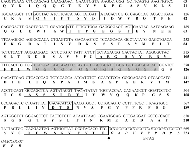
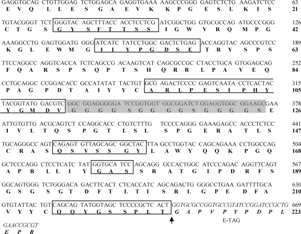
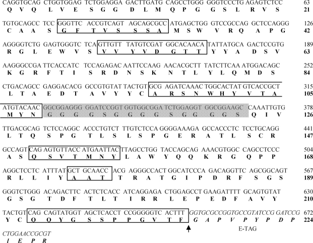
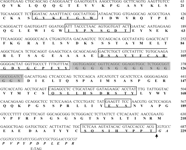
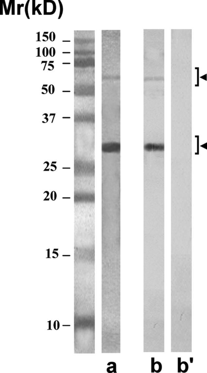
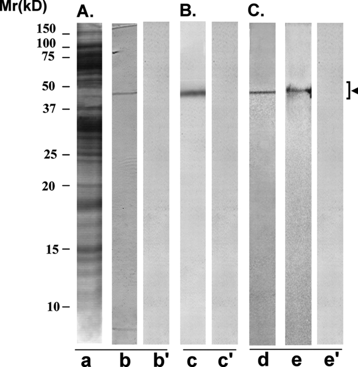
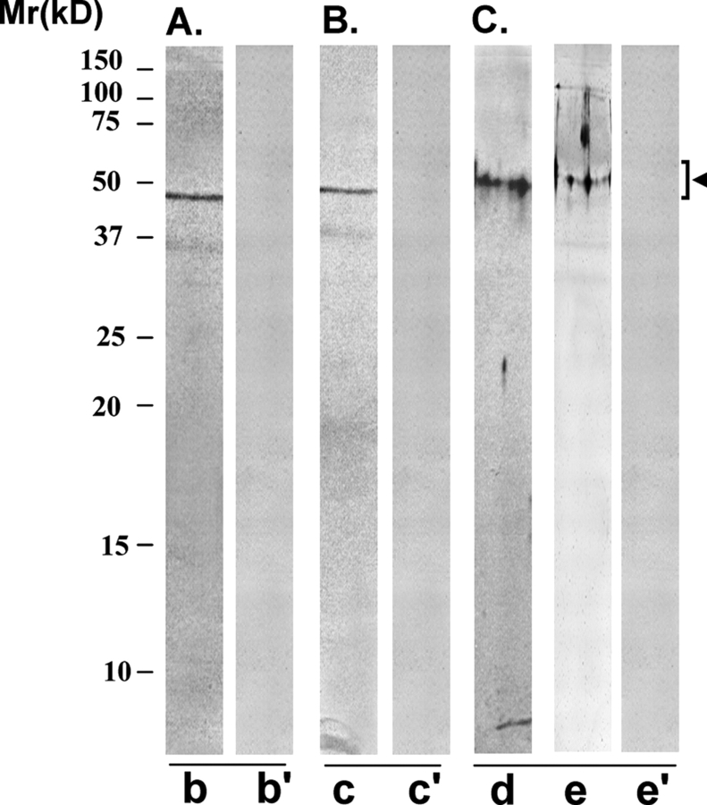
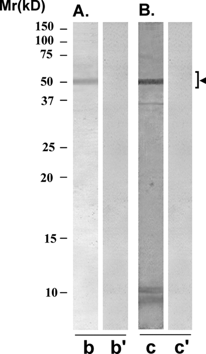
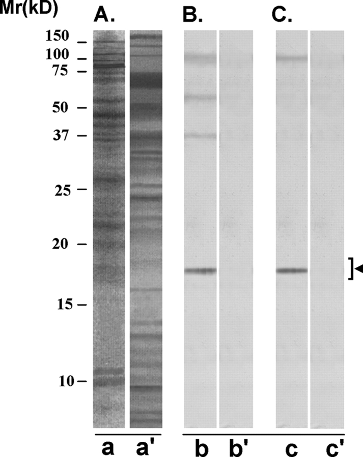
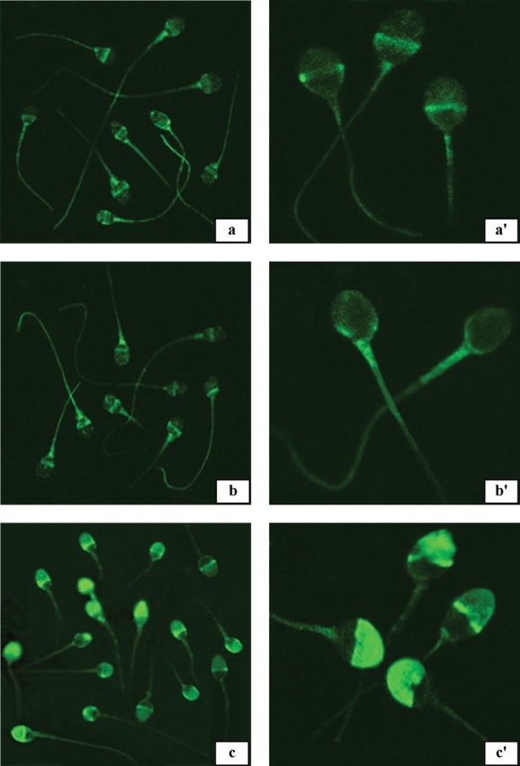
Results: Panning of the library against specific sperm antigens yielded several clones, and the four strongest reactive were selected for further analysis. These clones had novel sequences with unique complementarity-determining regions. ScFv antibodies were expressed, purified and analyzed for human sperm reactivity and effect on human sperm function. AFA-1 and FAB-7 scFv antibodies both reacted with fertilization antigen-1 antigen, but against different epitopes. YLP20 antibody reacted with the expected human sperm protein of 48 +/- 5 kDa. The fourth antibody, AS16, reacted with an 18 kDa sperm protein and seems to be a human homologue of the mouse monoclonal recombinant antisperm antibody that causes sperm agglutination. All these antibodies inhibited human sperm function.

Conclusions: This is the first study to report the use of phage display technology to obtain antisperm scFv antibodies of defined antigen specificity. These antibodies will find clinical applications in the development of novel immunocontraceptives, and specific diagnostics for immunoinfertility.


Figures

Figure 1:
Figure 1:
The cDNA sequence and the corresponding amino acid (aa) sequence of single chain variable fragment (scFv) clone (AFA-1) reactive with FA-1 antigen The aa sequence contains heavy chain (1–109 aa), linker (Gly4Ser)3 (110–124 aa) shown in shaded grey box, light chain (125–223 aa) and E-Tag (224–236 aa) sequence (shown after ↑ in italics). The aa sequence of immunoglobulin (Ig)G1 heavy chain includes framework region 1 (1–25 aa), framework region 2 (34–50 aa), framework region 3 (59–96 aa), and their corresponding CDR-1 (26–33 aa), CDR-2 (51–58 aa) and CDR-3 (97–109 aa) regions. Similarly, the aa sequence of Igк3 light chain includes framework region 1 (125–150 aa), framework region 2 (158–174 aa), framework region 3 (178–213 aa), and their corresponding CDR-1 (151–157 aa), CDR-2 (175–177 aa) and CDR-3 (214–223 aa) regions. All CDRs regions are enclosed in rectangular boxes.
Figure 2:
Figure 2:
The cDNA sequence and the corresponding amino acid (aa) sequence of scFv clone (FAB-7) reactive with FA1 antigen The aa sequence contains heavy chain (1–110 aa), linker (Gly4Ser)3 (111–125 aa) shown in shaded grey box, light chain (126–223 aa) and E-Tag (224–236 aa) sequence (shown after ↑ in italics). The aa sequence of IgG1 heavy chain includes framework region 1 (1–25 aa), framework region 2 (34–50 aa) and framework region 3 (58–95 aa), and their corresponding CDR-1 (26–33 aa), CDR-2 (51–57 aa) and CDR-3 (96–110 aa) regions. Similarly, the aa sequence of Igк3 light chain includes framework region 1 (126–151 aa), framework region 2 (159–175 aa) and framework region 3 (179–214 aa), and their corresponding CDR-1 (152–158 aa), CDR-2 (176–178 aa) and CDR-3 (215–223 aa) regions. All CDRs regions are enclosed in rectangular boxes.
Figure 3:
Figure 3:
The cDNA sequence and the corresponding amino acid (aa) sequence of scFv clone (YLP20) reactive with YLP12 antigen The aa sequence contains heavy chain (1–108 aa), linker (Gly4Ser)3 (109–123 aa) shown in shaded grey box, light chain (124–224 aa) and E-Tag (225–237 aa) sequence (shown after ↑ in italics). The aa sequence of IgG3 heavy chain includes framework region 1 (1–25 aa), framework region 2 (34–49 aa) and framework region 3 (58–95 aa), and their corresponding CDR-1 (26–33 aa), CDR-2 (50–57 aa) and CDR-3 (96–108 aa) regions. Similarly, the aa sequence of Igк3 light chain includes framework region 1 (123–149 aa), framework region 2 (157–173 aa) and framework region 3 (177–212 aa), and their corresponding CDR-1 (150–156 aa), CDR-2 (174–176 aa) and CDR-3 (213–224 aa) regions. All CDRs regions are enclosed in rectangular boxes.
Figure 4:
Figure 4:
The cDNA sequence and the corresponding amino acid (aa) sequence of scFv clone (AS16) reactive with HSE The aa sequence contains heavy chain (1–114 aa), linker (Gly4Ser)3 (115–129 aa) shown in shaded grey box, light chain (130–229 aa) and E-Tag (230–242 aa) sequence (shown after ↑ in italics). The aa sequence of IgG1 heavy chain includes framework region 1 (1–25 aa), framework region 2 (34–50 aa) and framework region 3 (59–96 aa), and their corresponding CDR-1 (26–33 aa), CDR-2 (51–58 aa) and CDR-3 (97–114 aa) regions. Similarly, the aa sequence of Igк2 light chain includes framework region 1 (130–153 aa), framework region 2 (165–181 aa) and framework region 3 (185–220 aa), and their corresponding CDR-1 (154–164 aa), CDR-2 (182–184 aa) and CDR-3 (221–229 aa) regions. All CDRs regions are enclosed in rectangular boxes.
Figure 5:
Figure 5:
Purity of the scFv antibodies isolated by using anti-E-Tag antibody column The purified scFv antibodies showed predominantly the expected single protein band of ∼28 kDa in SDS–PAGE after staining with silver nitrate (lane a). On freezing, the antibodies had a tendency to polymerize into dimeric form of ∼56 kDa, and sometimes into trimeric form of ∼84 kDa (not shown). All these forms were specifically recognized by the anti-E-Tag monoclonal antibody (lane b) and not by the myeloma control monoclonal antibody (lane b’) in the western blot procedure.
Figure 6:
Figure 6:
Immunoreactivity pattern of AFA-1 scFv antibody with HSE and FA-1 antigen The purified scFv antibody was examined for its immunoreactivity with LIS-solubilized HSE (A and B) and purified cognate human sperm FA-1 antigen (C), using western blot (A and C) and immunoprecipitation (B) procedures. (A) SDS–PAGE of HSE revealed several protein bands of various molecular identities after silver staining (lane a). AFA-1 scFv antibody specifically recognized a protein band of 50 ± 4 kDa, corresponding to FA-1 antigen on western blot of HSE (lane b). Control scFv antibody did not react with any specific band on the western blot (lane b’). (B) AFA-1 scFv antibody Sepharose 4B immunobeads reacted with a specific protein in HSE that on elution with glycine–HCl (0.1 M, pH 2.8) showed a single band of 50 ± 4 kDa, corresponding to FA-1 antigen, in SDS–PAGE (lane c). Control scFv antibody Sepharose 4B immunobeads did not react with any protein in HSE (lane c’). (C) The cognate FA-1 antigen purified from HSE using immunoaffinity column involving mouse monoclonal antibody MA-24 showed a single band of 50 ± 4 kDa in SDS–PAGE (lane d) that was specifically recognized by AFA-1 scFv antibody (lane e) and not by the control scFv antibody (lane e’) in the western blot procedure.
Figure 7:
Figure 7:
Immunoreactivity pattern of FAB-7 ScFv antibody with HSE and FA-1 antigen The purified scFv was examined for its reactivity with LIS-HSE (A and B) and purified cognate human sperm FA-1 antigen (C), using western blot (A and C) and immunoprecipitation (B) procedures. (A) FAB-7 scFv antibody specifically recognized a protein band of 50 ± 4 kDa, corresponding to FA-1 antigen, on western blot of HSE (lane b). Control scFv antibody did not react with any specific band on the western blot (lane b’). (B) FAB-7 scFv antibody Sepharose 4B immunobeads reacted with a specific protein in HSE that on elution with glycine–HCl (0.1 M, pH 2.8) showed a single band of 50 ± 4 kDa, corresponding to FA-1 antigen, in SDS–PAGE (lane c). Control scFv antibody Sepharose 4B immunobeads did not react with any protein in HSE (lane c’). (C) The cognate FA-1 antigen purified from HSE using immunoaffinity column involving mouse monoclonal antibody MA-24 showed a single band of 50 ± 4 kDa in SDS–PAGE (lane d), that was specifically recognized by FAB-7 ScFv antibody (lane e) and not by the control scFv antibody (lane e’) in the western blot procedure.
Figure 8:
Figure 8:
Immunoreactivity pattern of YLP20 scFv antibody with HSE The purified scFv was examined for its reactivity with LIS-solubilized HSE using western blot (A) and immunoprecipitation (B) procedures. (A) YLP20 scFv antibody specifically recognized a protein band of 48 ± 5 kDa, corresponding to YLP12 antigen, on western blot of HSE (lane b). Control scFv antibody did not react with any specific band on the western blot (lane b’). (B) YLP20 scFv antibody Sepharose 4B immunobeads reacted with a specific protein in HSE that on elution with glycine–HCl (0.1 M, pH 2.8) showed a single band of 48 ± 5 kDa in SDS–PAGE (lane c). Control scFv antibody Sepharose 4B immunobeads did not react with any protein in HSE (lane c’).
Figure 9:
Figure 9:
Immunoreactivity pattern of AS-16 scFv antibody with HSE The purified scFv antibody was examined for its reactivity with LIS-solubilized HSE (B) and MC-solubilized human sperm preparation (C), in the western blot procedure. (A) SDS–PAGE of LIS-solubilized human sperm preparation (lane a) and MC-solubilized human sperm preparation (lane a’) revealed several protein bands of various molecular identities after silver staining. (B) AS16 scFv antibody specifically recognized four protein bands of 18 (major band), 37, 55 and 100 kDa, respectively, on the western blot of LIS-solubilized human sperm preparation (lane b). Control scFv antibody did not react with any band on the western blot (lane b’). (C) AS-16 ScFv antibody recognized two protein bands of 18 (major band) and 100 kDa (minor band), in the western blot of MC-solubilized human sperm preparation (lane c). Control scFv antibody did not react with any specific band on the western blot (lane c’). The 18 kDa protein was the major band specifically recognized in both the LIS-solubilized human sperm preparation (lane a) and MC-solubilized human sperm preparation (lane a’) and it corresponds to SAGA-1 sperm protein.
Figure 10:
Figure 10:
Epifluorescent photomicrographs indicating the indirect immunofluorescent reaction pattern of AFA-1, FAB-7 and YLP20 scFv antibodies with methanol-fixed (a, b and c) and unfixed live human sperm (a', b', and c’) AFA-1 (a and a’) and FAB-7 (b and b’) antibodies predominantly reacted with post-acrosomal, midpiece and tail regions of methanol-fixed (a and b) and unfixed live human sperm (a' and b’). YLP20 antibody reacted with acrosomal, midpiece and tail regions of methanol-fixed (c) and unfixed live (c’) human sperm. Magnifications: a, b and c ×698; a', b' and c' ×1924.

Similar articles

Cited by

References

    1. Arndt KM, Muller KM, Pluckthun A. Factors influencing the dimer to monomer transition of an antibody single-chain Fv fragment. Biochemistry. 1998;37:12918–12926. - PubMed
    1. Bohring C, Krause W. Immune infertility: towards a better understanding of sperm (auto)-immunity. The value of proteomic analysis. Hum Reprod. 2003;18:915–924. - PubMed
    1. Byrd W, Tsu J, Wolf D. Kinetics of spontaneous and induced acrosomal loss in human sperm incubated under capacitating and noncapacitating conditions. Gamete Res. 1989;22:109–122. - PubMed
    1. Casadevall A. Passive antibody therapies: progress and continuing challenges. Clin Immunol. 1999;93:5–15. - PubMed
    1. Chapman AP. PEGylated antibodies and antibody fragments for improved therapy: a review. Adv Drug Deliv Rev. 2002;54:531–545. - PubMed

Publication types

Substances