Comparative Study
. 2008 May 15;586(10):2487-97.
doi: 10.1113/jphysiol.2007.148320. Epub 2008 Mar 27.

Brainstem modulation of locomotion in the neonatal mouse spinal cord

Affiliations
Comparative Study

Brainstem modulation of locomotion in the neonatal mouse spinal cord

Ian T Gordon et al. J Physiol. .

Abstract

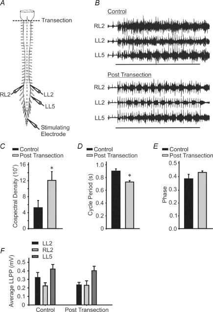
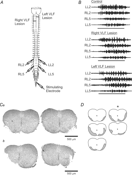
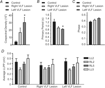
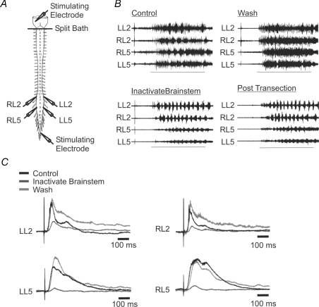
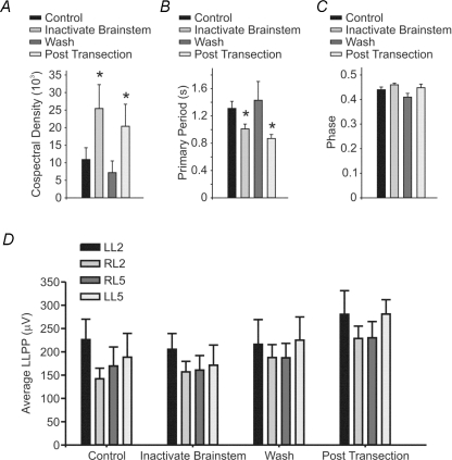
During development, descending projections to the spinal cord are immature. Available data suggest that even though these projections are not fully formed, they contribute to activation of spinal circuitry and promote development of network function. Here we examine the modulation of sacrocaudal afferent-evoked locomotor activity by descending pathways. We first examined the effects of brainstem transection on the afferent evoked locomotor-like rhythm using an isolated brainstem-spinal cord preparation of the mouse. Transection increased the frequency and stability of the locomotor-like rhythm while the phase remained unchanged. We then made histologically verified lesions of the ventrolateral funiculus and observed similar effects on the stability and frequency of the locomotor rhythm. We next tested whether these effects were due to downstream effects of the transection. A split-bath was constructed between the brainstem and spinal cord. Neural activity was suppressed in the brainstem compartment using cooled high sucrose solutions. This manipulation led to a reversible change in frequency and stability that mirrored our findings using lesion approaches. Our findings suggest that spontaneous brainstem activity contributes to the ongoing modulation of afferent-evoked locomotor patterns during early postnatal development. Our work suggests that some of the essential circuits necessary to modulate and control locomotion are at least partly functional before the onset of weight-bearing locomotion.


Figures

Figure 1
Figure 1. Brainstem transection modulates afferent-evoked rhythmicity
A, schematic diagram of the isolated brainstem–spinal cord showing recording and stimulation sites. The dotted line indicates the level of transection. B, representative traces illustrating the effect of brainstem transection on afferent-evoked rhythmicity. Horizontal lines represent the duration of the 10 s stimulus train. Stimulus trains were delivered every 3 min. CF, graphs show the cospectral density values (C), the cycle period (D), the phase (E) and the average LLPP (F) before and after transection. *Significant difference from control (n = 7, P < 0.05). Error bars represent s.e.m.
Figure 2
Figure 2. Discrete VLF lesions mimic the effect of complete brainstem transection
A, schematic diagram of the isolated brainstem–spinal cord showing recording and stimulation sites. Horizontal lines represent the levels of the two lesion sites. B, representative traces illustrating the effects of VLF lesions on afferent-evoked rhythmicity. The right VLF trace represents a right ipsilateral lesion. The left VLF trace represents activity after both ipsilateral and contralateral sides were lesioned. Horizontal lines represent the duration of the 10 s stimulus train. C, haematoxylin- and eosin-stained 6 μm cervical spinal cord slices showing the left and right lesion sites for the cords with the smallest (a) and largest (b) lesions. D, schematic diagram of the lesion sizes and locations for each of the five cords in the experimental set. Shaded grey areas represent the lesioned portions of the spinal cord. The cord with an asterisk represents that shown in B.
Figure 3
Figure 3. The cospectral density and primary period of the afferent-evoked rhythmicity are both altered by discrete VLF lesions
Graphs representing the mean cospectral density (A), primary period (B), phase (C) and average LLPP (D) under control conditions and after one side and then after both sides have received a lesion of the VLF. *Significant difference from control (n = 5, P < 0.05). Error bars represent s.e.m.
Figure 4
Figure 4. Brainstem inactivation mimics the effect of complete brainstem transection
A, schematic diagram of the isolated brainstem–spinal cord showing recording and stimulation sites and the location of the split bath, represented by a horizontal line. B, representative traces illustrating the effects of brainstem inactivation and brainstem transection on afferent-evoked rhythmicity. Horizontal lines represent the duration of the 10 s stimulus train. C, representative traces illustrating the effects of brainstem inactivation on ventral root potentials evoked by single brainstem stimulating pulses.
Figure 5
Figure 5. The cospectral density and primary period of the afferent-evoked rhythmicity are both altered by brainstem inactivation
Graphs representing the cospectral density (A), primary period (B), phase (C) and average LLPP (D) under control conditions, after brainstem inactivation via cooling to below 10°C and the circulation of a high sucrose solution, after washout and reheating and finally after brainstem transection. *Significant difference from control (n = 8, P < 0.05). Error bars represent s.e.m.

Similar articles

Cited by

References

    1. Atsuta Y, Garcia-Rill E, Skinner RD. Characteristics of electrically induced locomotion in rat in vitro brain stem-spinal cord preparation. J Neurophysiol. 1990;64:727–735. - PubMed
    1. Beato M, Nistri A. Serotonin-induced inhibition of locomotor rhythm of the rat isolated spinal cord is mediated by the 5-HT1 receptor class. Proc Biol Sci. 1998;265:2073–2080. - PMC - PubMed
    1. Blivis D, Mentis GZ, O'Donovan MJ, Lev-Tov A. Differential effects of opioids on sacrocaudal afferent pathways and central pattern generators in the neonatal rat spinal cord. J Neurophysiol. 2007;97:2875–2886. - PubMed
    1. Cazalets JR, Sqalli-Houssaini Y, Clarac F. Activation of the central pattern generators for locomotion by serotonin and excitatory amino acids in neonatal rat. J Physiol. 1992;455:187–204. - PMC - PubMed
    1. Cowley KC, Schmidt BJ. A comparison of motor patterns induced by N-methyl-D-aspartate, acetylcholine and serotonin in the in vitro neonatal rat spinal cord. Neurosci Lett. 1994;171:147–150. - PubMed

Publication types

LinkOut - more resources