Comparative Study
. 2008 May 15;586(10):2523-38.
doi: 10.1113/jphysiol.2007.148197. Epub 2008 Mar 27.

Selective regulation of spontaneous activity of neurons of the deep cerebellar nuclei by N-type calcium channels in juvenile rats

Affiliations
Comparative Study

Selective regulation of spontaneous activity of neurons of the deep cerebellar nuclei by N-type calcium channels in juvenile rats

Karina Alviña et al. J Physiol. .

Abstract

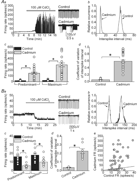
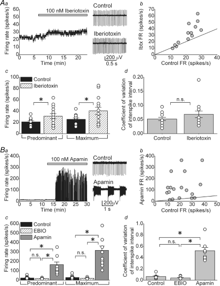
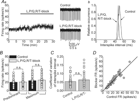
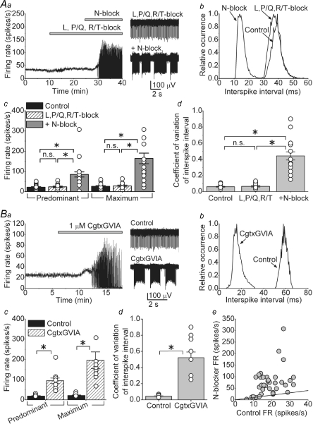
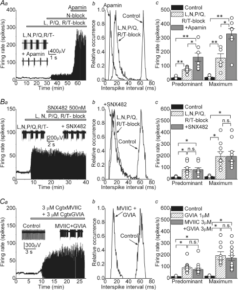
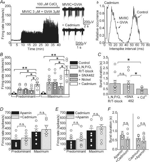
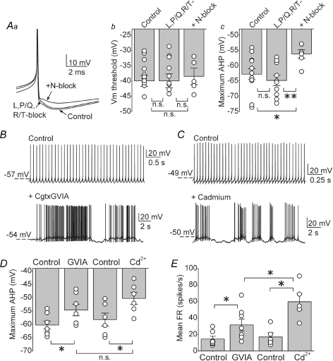
The cerebellum coordinates movement and maintains body posture. The main output of the cerebellum is formed by three deep nuclei, which receive direct inhibitory inputs from cerebellar Purkinje cells, and excitatory collaterals from mossy and climbing fibres. Neurons of deep cerebellar nuclei (DCN) are spontaneously active, and disrupting their activity results in severe cerebellar ataxia. It is suggested that voltage-gated calcium channels make a significant contribution to the spontaneous activity of DCN neurons, although the exact identity of these channels is not known. We sought to delineate the functional role and identity of calcium channels that contribute to pacemaking in DCN neurons of juvenile rats. We found that in the majority of cells blockade of calcium currents results in avid high-frequency bursting, consistent with the notion that the net calcium-dependent current in DCN neurons is outward. We showed that the bursting seen in these neurons after block of calcium channels is the consequence of reduced activation of small-conductance calcium-activated (SK) potassium channels. With the use of selective pharmacological blockers we showed that L-, P/Q-, R- and T-type calcium channels do not contribute to the spontaneous activity of DCN neurons. In contrast, blockade of high-threshold N-type calcium channels increased the firing rate and caused the cells to burst. Our results thus suggest a selective coupling of N-type voltage-gated calcium channels with calcium-activated potassium channels in DCN neurons. In addition, we demonstrate the presence of a cadmium-sensitive calcium conductance coupled with SK channels, that is pharmacologically distinct from L-, N-, P/Q-, R- and T-type calcium channels.


Figures

Figure 1
Figure 1. Cadmium-sensitive calcium channels make a significant contribution to the spontaneous activity of the majority of DCN neurons
Spontaneous activity of individual, visually identified DCN neurons was monitored by extracellular recording in acutely prepared slices at 35°C. Fast excitatory and inhibitory synaptic transmission was blocked pharmacologically. Aa, application of cadmium increased the firing rate and caused random bursts of action potentials in the majority of DCN neurons. Sample raw data traces are shown in the right panel. Ab, interspike interval histogram of the cell shown in Aa. Ac, predominant and maximum spontaneous firing rates of individual cells together with the averages in the presence and absence of cadmium. *P < 0.001, n = 33 cells. Ad, the coefficient of variation of the interspike intervals for the same cells shown in Ac. *P < 0.001. Ba, in a small fraction of cells cadmium simply reduced the firing rate. Sample traces are shown to the right of the panel. Bb, interspike interval histogram of the cell shown in Ba. Bc, predominant and maximum spontaneous firing rates of individual cells together with the averages in the presence and absence of cadmium. *P < 0.01, n = 11 cells. Bd, the coefficient of variation of the interspike intervals for the same cells shown in Bc. *P < 0.001. Be, the predominant firing rate of all cells examined (in A and B) before and after addition of cadmium. The continuous line represents the unity line.
Figure 2
Figure 2. KCa channels, particularly of the SK type, control the rate of intrinsic activity of DCN neurons
Aa, the spontaneous activity of a DCN neuron as large-conductance KCa (BK) channels were blocked with 100 nm iberiotoxin. Ab, the firing rate of DCN neurons before (control) and after application of iberiotoxin. The continuous line denotes unity. Ac, predominant and maximum spontaneous firing rates of individual cells together with the averages in the presence and absence of iberiotoxin. *P < 0.05, n = 14 cells. Ad, the coefficient of variation of the interspike intervals for the same cells shown in Ac. Ba, blockade of small-conductance (SK) KCa channels with 100 nm apamin increased the firing rate and resulted in high-frequency bursts of action potentials. Bb, the firing rate of DCN neurons before (control) and after application of apamin. The continuous line denotes unity. Bc, predominant and maximum spontaneous firing rates of individual cells together with the averages in the presence and absence of 20 μm EBIO (n = 7) or 100 nm apamin (n = 14). *P < 0.001. Bd, the coefficient of variation of the interspike intervals for the same cells shown in Bc. *P < 0.001. n.s., not significant.
Figure 3
Figure 3. L-, P/Q- and R/T-type voltage-gated calcium channels do not contribute to the spontaneous activity of DCN neurons
A, bath application of a combination of L-, P/Q- and R/T-calcium channels blockers (2 μm nimodipine, 200 nm ω-agatoxin IVA and 5 μm mibefradil, respectively) did not significantly alter the spontaneous activity of a DCN neuron. Ab, interspike interval histogram of the cell shown in A. B, predominant and maximum spontaneous firing rates of individual cells together with the averages in the presence and absence of a combination of L-, R/T- and P/Q-type calcium channel blockers. n = 50 cells. C, the coefficient of variation of the interspike intervals for the same cells shown in B. D, the firing rate of all cells examined before and after addition of L-, R/T- and P/Q-type calcium channel blockers. The continuous line represents the unity line. n.s., not significant.
Figure 4
Figure 4. Block of N-type calcium channels increases the firing rate and causes bursting in the majority of DCN neurons
Aa, the rate of spontaneous activity of a DCN neuron in control solution and after sequential addition of a mixture of L-, P/Q- and R/T-type calcium channel blockers (2 μm nimodipine, 200 nm ω-agatoxin IVA and 5 μm mibefradil) and 1 μm ω-conotoxin (Cgtx) GVIA to block N-type calcium channels. Representative raw data traces are also shown. Ab, interspike interval histogram of the cell shown in Aa. Ac, predominant and maximum spontaneous firing rates of individual cells together with the averages in the presence and absence of the mixture of L-, P/Q- and R/T-type calcium channel blockers, and after the subsequent addition of a N-type calcium channel blocker (either 1 μm Cgtx GVIA or 1 μm Cgtx MVIIC). *P < 0.001; n.s., not significant, n = 22 cells. Ad, the coefficient of variation of the interspike intervals for the same cells shown in Ac. *P < 0.001; n.s., not significant. Ba, application of 1 μm Cgtx GVIA to block N-type calcium channels in the absence of any other calcium channel blocker also increased the firing rate and caused the cell to burst. Bb, interspike interval histogram of the cell shown in Ba. Bc, predominant and maximum spontaneous firing rates of individual cells together with the averages in the presence and absence of Cgtx GVIA. *P < 0.001, n = 14 cells. Bd, the coefficient of variation of the interspike intervals for the same cells shown in Bc. *P < 0.001. Be, the firing rate of all cells examined (in A and B) before and after the addition of the N-type calcium channel blocker. The continuous line represents the unity line.
Figure 5
Figure 5. Additional calcium influx pathways activate SK channels and contribute to the firing rate of DCN neurons
Aa, the rate of spontaneous activity of a DCN neuron in control solution and after sequential addition of a mixture of L-, P/Q- and R/T-type calcium channel blockers (2 μm nimodipine, 200 nm ω-agatoxin IVA and 5 μm mibefradil), then 1 μm Cgtx GVIA to block N-type calcium channels, and 100 nm apamin to block SK channels. The inset shows raw traces before and after application of apamin. Ab, interspike interval histogram of the cell shown in Aa. Ac, predominant and maximum spontaneous firing rates of individual cells together with the averages in control, after L-, N-, P/Q- and R/T-type calcium channels were blocked by 2 μm nimodipine, 1 μm Cgtx GVIA, 200 nm ω-agatoxin IVA and 5 μm mibefradil (L-, N-, P/Q- and R/T-block), and after the successive addition of 100 nm apamin. *P < 0.01 in predominant FR and P < 0.05 in maximum FR. **P < 0.001, n = 8 cells. Ba, the rate of spontaneous activity of a DCN neuron in control solution and after sequential addition of L-, N-, P/Q- and R/T-type calcium channels blockers, and 500 nm SNX-482 to specifically block R-type calcium channels. The inset shows example of raw traces obtained before and after addition of SNX-482. Bb, histogram of the interspike interval distribution of the cell in Ba. Bc, individual and average firing rates obtained in 9 cells in control, after blocking L-, N-, P/Q- and R/T-type calcium channels and after blocking all R-type channels with SNX-482 *P < 0.001. Ca, continuous recording of a cell in control conditions and after adding a combination of Cgtx MVIIC and Cgtx GVIA both at 3 μm. The inset shows example of raw data showing the bursts observed after the toxins. Cb, distribution of interspike intervals before and after concurrent addition of 3 μm Cgtx MVIIC and 3 μm Cgtx GVIA. Cc, average and individual data from the cells exposed to the combination of toxins before and after. The FR obtained in the presence of 1 μm Cgtx GVIA is also plotted to allow direct comparison. *P < 0.001; n.s., not significant, n = 17.
Figure 6
Figure 6. Blockade of calcium influx with cadmium has comparable effects as block of SK channels with apamin
Aa, application of 100 μm cadmium after concurrent application of Cgtx MVIIC and GVIA both at 3 μm further increased the firing rate and reduced the duration of the bursts. Sample raw data are also shown. Ab, histogram of the interspike interval of the cell in Aa. B, average and individual data obtained after sequentially adding: a mixture of blockers of L-, N-, P/Q- and R/T-type calcium channels (2 μm nimodipine, 1 μm Cgtx GVIA, 200 nm ω-agatoxin and 5 μm mibefradil), then 500 nm SNX-482 (to block R-type calcium channels), then 50 μm nickel and finally 100 μm cadmium. *P < 0.05, **P < 0.001, n = 9 cells. C, average and individual data showing the duration of the bursts for the same cells presented in Fig. 6B. *P < 0.05. D, average and individual firing rates in cells first exposed to 100 nm apamin and then to 100 μm cadmium. E, average and individual firing rates in cells first exposed to 100 μm cadmium and then to 100 nm apamin. F, average and individual data showing the duration of the bursts for the same cells presented in D and E. n.s., not significant.
Figure 7
Figure 7. N-type, but not L-, P/Q- or R/T-type, calcium channels significantly contribute to the action potential afterhyperpolarization in DCN neurons
Aa, truncated traces of action potentials obtained from whole-cell current clamp recording from a DCN neuron in I = 0 mode before (control), and after sequential additions of a mixture of L-, P/Q- and R/T-type calcium channel blockers (2 μm nimodipine, 200 nm ω-agatoxin IVA and 5 μm mibefradil) and 1 μm Cgtx GVIA to block N-type calcium channels. Ab, the action potential threshold of individual cells together with the averages in control solution, in the presence of a mixture of L-, P/Q- and R/T-type calcium channel blockers (2 μm nimodipine, 200 nm ω-agatoxin IVA and 5 μm mibefradil), and subsequent block of N-type calcium channels with 1 μm Cgtx GVIA. Ac, the average maximum afterhyperpolarization (AHP) and individual data for the same cells and conditions shown in B. *P < 0.03, **P < 0.005. n = 12 cells for control and L-, P/Q- and R/T-type calcium channels, n = 6 after addition of N-type blocker. B, raw traces of whole-cell current clamp recordings in control conditions and after adding 1 μm Cgtx GVIA. C, similar to B; current clamp recordings before and after application of 100 μm cadmium. D, maximum AHP of the action potential waveform measured in the cells recorded soon after establishing whole-cell configuration (B and C). Average and individual data are plotted per condition. *P < 0.01. n = 7 cells in Cgtx GVIA and n = 6 cells in cadmium. E, mean firing rate of the same cells in D. The graph shows the average and the individual values obtained in each condition. *P < 0.05. n.s., not significant.

Similar articles

Cited by

References

    1. Aizenman CD, Linden DJ. Regulation of the rebound depolarization and spontaneous firing patterns of deep nuclear neurons in slices of rat cerebellum. J Neurophysiol. 1999;82:1697–1709. - PubMed
    1. Blatz AL, Magleby KL. Single apamin-blocked Ca-activated K+ channels of small conductance in cultured rat skeletal muscle. Nature. 1986;323:718–720. - PubMed
    1. Bloodgood BL, Sabatini BL. Nonlinear regulation of unitary synaptic signals by CaV2.3 voltage-sensitive calcium channels located in dendritic spines. Neuron. 2007;53:249–260. - PubMed
    1. Boland LM, Morrill JA, Bean BP. omega-Conotoxin block of N-type calcium channels in frog and rat sympathetic neurons. J Neurosci. 1994;14:5011–5027. - PMC - PubMed
    1. Bond CT, Sprengel R, Bissonnette JM, Kaufmann WA, Pribnow D, Neelands T, et al. Respiration and parturition affected by conditional overexpression of the Ca2+-activated K+ channel subunit, SK3. Science. 2000;289:1942–1946. - PubMed

Publication types

Substances

LinkOut - more resources