. 2008 Jul 1;17(13):2018-29.
doi: 10.1093/hmg/ddn099. Epub 2008 Mar 27.

Diverse driving forces underlie the invariant occurrence of the T42A, E139D, I282V and T468M SHP2 amino acid substitutions causing Noonan and LEOPARD syndromes

Affiliations

Diverse driving forces underlie the invariant occurrence of the T42A, E139D, I282V and T468M SHP2 amino acid substitutions causing Noonan and LEOPARD syndromes

Simone Martinelli et al. Hum Mol Genet. .

Abstract

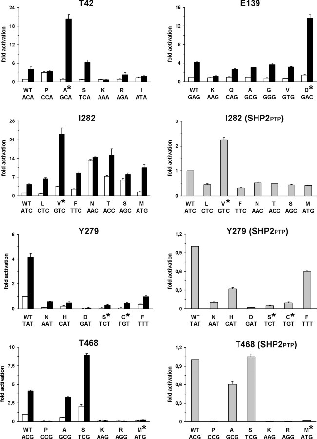
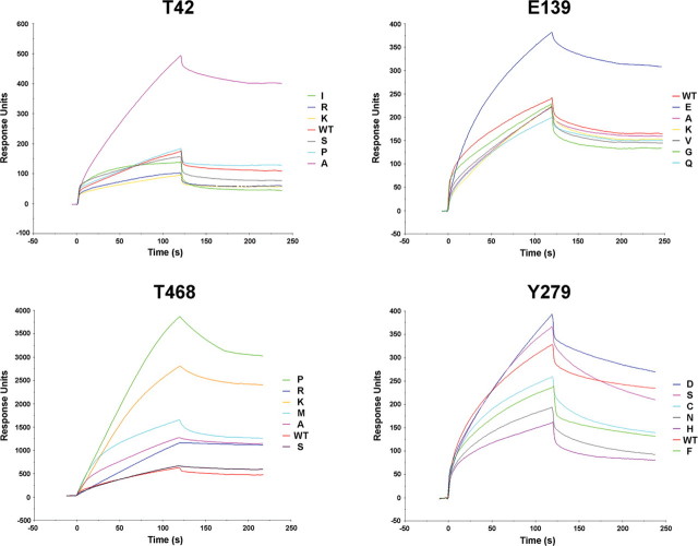
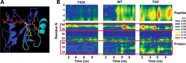
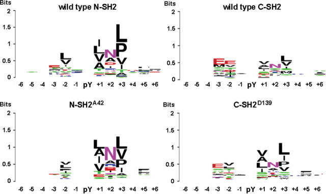
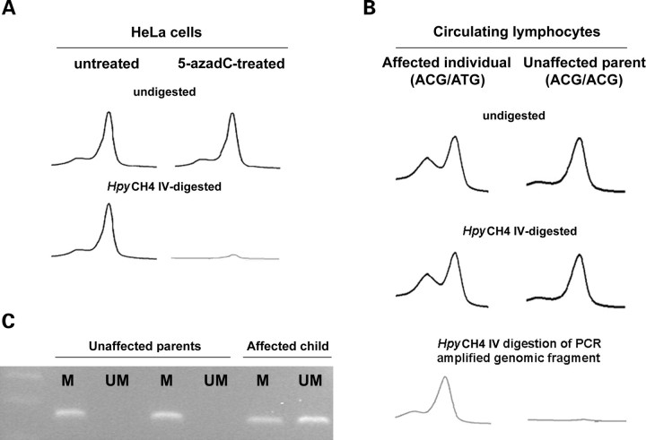
Missense PTPN11 mutations cause Noonan and LEOPARD syndromes (NS and LS), two developmental disorders with pleiomorphic phenotypes. PTPN11 encodes SHP2, an SH2 domain-containing protein tyrosine phosphatase functioning as a signal transducer. Generally, different substitutions of a particular amino acid residue are observed in these diseases, indicating that the crucial factor is the residue being replaced. For a few codons, only one substitution is observed, suggesting the possibility of specific roles for the residue introduced. We analyzed the biochemical behavior and ligand-binding properties of all possible substitutions arising from single-base changes affecting codons 42, 139, 279, 282 and 468 to investigate the mechanisms underlying the invariant occurrence of the T42A, E139D and I282V substitutions in NS and the Y279C and T468M changes in LS. Our data demonstrate that the isoleucine-to-valine change at codon 282 is the only substitution at that position perturbing the stability of SHP2's closed conformation without impairing catalysis, while the threonine-to-alanine change at codon 42, but not other substitutions of that residue, promotes increased phosphopeptide-binding affinity. The recognition specificity of the C-SH2 domain bearing the E139D substitution differed substantially from its wild-type counterpart acquiring binding properties similar to those observed for the N-SH2 domain, revealing a novel mechanism of SHP2's functional dysregulation. Finally, while functional selection does not seem to occur for the substitutions at codons 279 and 468, we point to deamination of the methylated cytosine at nucleotide 1403 as the driving factor leading to the high prevalence of the T468M change in LS.


Figures

Figure 1.
Figure 1.
Biochemical characterization of SHP2 mutants. In vitro phosphatase assay of wild-type SHP2 and all possible mutants arising from a single-base change at codons 42, 139, 282, 279 and 468. Catalytic activity was measured as pmoles of phosphate released using pNPP as substrate, basally (white bars) and following stimulation with the PTPNS1 BTAM peptide (black bars). Activities of recombinant wild-type SHP2PTP encoding for the isolated PTP domain and mutants at positions 282, 279 and 468 are also reported (gray bars). Values are expressed as mean ± standard deviation of at least three independent experiments and are normalized to the basal activity of the wild-type enzyme.
Figure 2.
Figure 2.
SPR analysis. Sensorgrams of the interaction of recombinant wild-type SHP2 or mutants at codons 42, 139, 279 and 468 with the biotinylated PTPNS1 BTAM peptide. Following binding of the phosphopeptide over a streptavidin-coated sensor chip, recombinant proteins were applied, and the amount of interacting protein was monitored by the change in arbitrary response units as a function of time at 25°C.
Figure 3.
Figure 3.
MD simulations. (A) Structure of the N-SH2 domain complexed with the PDGFRB SVLpYTAVQP phosphopeptide (pdb entry 1AYA). The peptide is shown in red with the N-terminal residues (−3 to −1) and the phosphorylated tyrosine (pY) highlighted in orange and yellow, respectively. The N-SH2 domain is displayed in blue with the BC loop, N-terminal helix and adjacent loop highlighted in cyan. The side chain of T42 is shown in green. (B) Mobility of Cα atoms in selected protein and phosphopeptide regions during the simulations of wild-type SHP2, SHP2A42 and SHP2I42. Plots report the atom's root mean square fluctuations as a function of time and residue number. Phosphopeptide Cα atoms are numbered with respect to the pY residue (indicated as 0). Red boxes highlight protein regions where significant mobility differences are observed, corresponding to the BC loop (residues 34–40), the N-terminal helix (residues 13–22) and the loop preceding it (residues 9–12).
Figure 4.
Figure 4.
WebLogo representation of binding recognition specificity of SHP2’s SH2 domains. Sequence logo representation generated for the wild-type N-SH2 and C-SH2 domains (above) and derived mutants bearing the T42A or E139D substitution (below). Each logo abridges the residue enrichment at the positions flanking the phosphorylated tyrosine (pY) in the best ligand peptides and was obtained by comparing the amino acid recurrence of bound peptides with a signal higher than average plus 1 standard deviation. For each position, the overall stack height indicates the sequence conservation, while the height of symbols within each stack indicates the relative frequency of the indicated residue. Residue positions are numbered starting from the pY (indicated as 0).
Figure 5.
Figure 5.
CpG methylation status at codon 468. (A) DHPLC profiles showing missing of PTPN11 exon 12 PCR product in HeLa cells treated with the DNA demethylating agent 5-azadC and digested with the methylation-sensitive enzyme HpyCH4 IV. (B) DHPLC profiles showing amplification of PTPN11 exon 12 from genomic DNA obtained from peripheral lymphocites of a subject heterozygous for the LS-causing C1403T mutation and unaffected parents, digested or not with the HpyCH4 IV endonuclease. Efficiency of the treatment was confirmed by digestion of an amplified genomic fragment encompassing exon 12 and subsequent re-amplification of the digested product. (C) Methylation-specific PCR assay performed on genomic DNA obtained from the same members of the family. Amplification was carried out using primers opportunely designed to amplify either the methylated (M) or the unmethylated (UM) alleles.

Similar articles

Cited by

References

    1. Tartaglia M., Mehler E.L., Goldberg R., Zampino G., Brunner H.G., Kremer H., van der Burgt I., Crosby A.H., Ion A., Jeffery S., et al. Mutations in PTPN11, encoding the protein tyrosine phosphatase SHP2, cause Noonan syndrome. Nat. Genet. 2001;29:465–468. - PubMed
    1. Tartaglia M., Kalidas K., Shaw A., Song X., Musat D.L., van der Burgt I., Brunner H.G., Bertola D.R., Crosby A., Ion A., et al. PTPN11 mutations in Noonan syndrome: molecular spectrum, genotype-phenotype correlation, and phenotypic heterogeneity. Am. J. Hum. Genet. 2002;70:1555–1563. - PMC - PubMed
    1. Digilio M.C., Conti E., Sarkozy A., Mingarelli R., Dottorini T., Marino B., Pizzuti A., Dallapiccola B. Grouping of multiple-lentigines/ LEOPARD and Noonan syndromes on the PTPN11 gene. Am. J. Hum. Genet. 2002;71:389–394. - PMC - PubMed
    1. Legius E., Schrander-Stumpel C., Schollen E., Pulles-Heintzberger C., Gewillig M., Fryns J.P. PTPN11 mutations in LEOPARD syndrome. J. Med. Genet. 2002;39:571–574. - PMC - PubMed
    1. Noonan J. Hypertelorism with Turner phenotype. A new syndrome with associated congenital heart disease. Am. J. Dis. Child. 1968;116:373–380. - PubMed

Publication types

MeSH terms

Substances