. 2008 Jul;149(7):3361-9.
doi: 10.1210/en.2008-0133. Epub 2008 Mar 27.

Estrogen inhibits cardiac hypertrophy: role of estrogen receptor-beta to inhibit calcineurin

Affiliations

Estrogen inhibits cardiac hypertrophy: role of estrogen receptor-beta to inhibit calcineurin

Ali Pedram et al. Endocrinology. 2008 Jul.

Abstract

Estrogen has been reported to prevent development of cardiac hypertrophy in female rodent models and in humans. However, the mechanisms of sex steroid action are incompletely understood. We determined the cellular effects by which 17beta-estradiol (E2) inhibits angiotensin II (AngII)-induced cardiac hypertrophy in vivo. Two weeks of angiotensin infusion in female mice resulted in marked hypertrophy of the left ventricle, exacerbated by the loss of ovarian steroid hormones from oophorectomy. Hypertrophy was 51% reversed by the administration of E2 (insertion of 0.1 mg/21-d-release tablets). The effects of E2 were mainly mediated by the estrogen receptor (ER) beta-isoform, because E2 had little effect in ERbeta-null mice but comparably inhibited AngII-induced hypertrophy in wild-type or ERalpha-null mice. AngII induced a switch of myosin heavy chain production from alpha to beta, but this was inhibited by E2 via ERbeta. AngII-induced ERK activation was also inhibited by E2 through the beta-receptor. E2 stimulated brain natriuretic peptide protein expression and substantially prevented ventricular interstitial cardiac fibrosis (collagen deposition) as induced by AngII. Importantly, E2 inhibited calcineurin activity that was stimulated by AngII, related to E2 stimulating the modulatory calcineurin-interacting protein (MCIP) 1 gene and protein expression. E2 acting mainly through ERbeta mitigates the important signaling by AngII that produces cardiac hypertrophy and fibrosis in female mice.


Figures

Figure 1
Figure 1
E2 prevents cardiac hypertrophy induced by AngII. A, Whole hearts were removed and weighed for calculating the ratio of heart weight to body weight. The bar graph is the mean ± sem from six to eight mice per condition. *, P < 0.05 for WT vs. same receiving AngII or ovariectomized (Ovx) WT vs. same plus AngII. +, P < 0.05 for Ovx WT plus AngII vs. same plus E2. Mice receiving E2 alone were similar to control mice (data not shown). Bar, 2 mm. B, Transverse sections of the ventricles stained with hematoxylin and eosin. a–c, WT mice; d and e, ERαKO mice; f and g, ERβKO mice; a is saline-infused control; b, d, and f are AngII infusion; and c, e, and g are AngII plus E2 replacement. All mice were ovariectomized. The bar graph represents six to eight mice per condition. *, P < 0.05 for WT (saline control) vs. other condition; +, P < 0.05 for AngII vs. same plus E2 for WT or ERαKO mouse. Bar, 2 mm.
Figure 2
Figure 2
E2 prevents AngII-induced cardiac fibrosis. A, Representative Masson trichrome staining of collagen deposition in the left ventricle of ovariectomized female mice exposed to the indicated conditions is shown (n = 5 mice per condition). Arrows indicate fibrosis. Bar, 0.1 mm. B, The percent area of fibrosis is quantified as described in Materials and Methods, and the bar graph data are the means ± sem (n = 5 mice per condition). C, Hydroxyproline content of the ventricle was measured by spectrophotometry, and means ± sem were calculated. *, P < 0.05 for control vs. AngII or vs. AngII plus E2 in the ERβKO mice; +, P < 0.05 for AngII vs. AngII plus E2.
Figure 3
Figure 3
E2 modulates important markers of cardiac hypertrophy. A, MHCα and -β proteins were determined by immunoblot from the pooled ventricles of six mice per condition. GAPDH is shown as a loading control. B, BNP expression in the left ventricle of mice. Individual mouse ventricles were processed for protein extraction to determine BNP expression by immunoblot (n = 6 per condition). *, P < 0.05 for control vs. AngII or E2 or both together. C, AngII activation of ERK is inhibited by E2. Kinase activity in the left ventricle was determined at 2 wk of treatment, from equal amounts of ERK2 protein immunoprecipitated from the three mouse models. Total ERK2 protein is shown as loading control. MBP is myelin basic protein substrate. Bar graph is the mean ± sem of three experiments combined. *, P < 0.05 for control vs. AngII or AngII plus E2 in ERβKO mice; +, P < 0.05 for AngII vs. AngII plus E2.
Figure 4
Figure 4
Inhibition of calcineurin and stimulation of MCIP1 by E2. A, Calcineurin activity was determined in individual samples from six mouse ventricles per condition in WT or KO mice. *, P < 0.05 for control vs. AngII; +, P < 0.05 for AngII vs. AngII plus E2. B, MCIP gene and protein expression were determined by PCR (top panel) from the pooled ventricular samples of mice (n = 6 per condition) and by Western blot (bottom panel), respectively. Actin is shown as loading control.
Figure 5
Figure 5
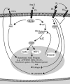
E2 and ERβ inhibit AngII-induced cardiac hypertrophy. The illustration represents data from in vitro (17) and in vivo studies here. AngII binds the AT1 receptor that activates Gqα and Gβγ signaling to up-regulate calcium. This signaling induces the activity of protein phosphatase 2B (calcineurin), dephosphorylating NFAT transcription factors in the cytosol. As a result, NFAT proteins (especially NFATc3) move to the nucleus where they collaborate with GATA-4 and MEF-2 transcription factors to stimulate the hypertrophic gene program. AngII also stimulates ERK MAPK signaling to effect gene up-regulation. E2 acting through ERβ stimulates the MCIP1 gene via PI3 kinase (PI3K) signaling, the protein product of which binds/clamps the catalytic activity of calcineurin. This prevents NFAT translocation to the nucleus and inhibits the hypertrophic genes required for cardiomyocyte increase in size (hypertrophy). E2 activating both ERα and ERβ stimulates the ANP and BNP genes, whose secreted protein products bind the guanylate cyclase A receptor and inhibit AngII-induced ERK activation. NCX, Sodium/calcium exchanger.

Similar articles

Cited by

References

    1. Kim J, Levin ER 2006 Estrogen signaling in the cardiovascular system. Nuclear Receptor Signaling 4:e013–e017 - PMC - PubMed
    1. Dunn FG, Pfeffer MA 1999 Left ventricular hypertrophy in hypertension. N Engl J Med 340:1279–1280 - PubMed
    1. Saxon LA, De Marco T 2002 Arrhythmias associated with dilated cardiomyopathy. Card Electrophysiol Rev 6:18–25 - PubMed
    1. Wagenaar LJ, Voors AA, Buikema H, van Gilst WH 2002 Angiotensin receptors in the cardiovascular system. Can J Cardiol 18:1331–1339 - PubMed
    1. D'Angelo DD, Sakata Y, Lorenz JN, Boivin GP, Walsh RA, Liggett SB, Dorn II GW 1997 Transgenic Gαq overexpression induces cardiac contractile failure in mice. Proc Natl Acad Sci USA 94:8121–8126 - PMC - PubMed

Publication types

MeSH terms