. 2008 Jun;83(6):1512-21.
doi: 10.1189/jlb.0108076. Epub 2008 Apr 17.

Inhibition of corneal inflammation by liposomal delivery of short-chain, C-6 ceramide

Affiliations

Inhibition of corneal inflammation by liposomal delivery of short-chain, C-6 ceramide

Yan Sun et al. J Leukoc Biol. 2008 Jun.

Abstract

Ceramide is recognized as an antiproliferative and proapoptotic sphingolipid metabolite; however, the role of ceramide in inflammation is not well understood. To determine the role of C6-ceramide in regulating inflammatory responses, human corneal epithelial cells were treated with C6-ceramide in 80 nm diameter nanoliposome bilayer formulation (Lip-C6) prior to stimulation with UV-killed Staphylococcus aureus. Lip-C6 (5 muM) inhibited the phosphorylation of proinflammatory and proapoptotic MAP kinases JNK and p38 and production of neutrophil chemotactic cytokines CXCL1, CXCL5, and CXCL8. Lip-C6 also blocked CXC chemokine production by human and murine neutrophils. To determine the effect of Lip-C6 in vivo, a murine model of corneal inflammation was used in which LPS or S. aureus added to the abraded corneal surface induces neutrophil infiltration to the corneal stroma, resulting in increased corneal haze. Mice were treated topically with 2 nMoles (811 ng) Lip-C6 or with control liposomes prior to, or following, LPS or S. aureus stimulation. We found that corneal inflammation was significantly inhibited by Lip-C6 but not control liposomes given prior to, or following, activation by LPS or S. aureus. Furthermore, Lip-C6 did not induce apoptosis of corneal epithelial cells in vitro or in vivo, nor did it inhibit corneal wound healing. Together, these findings demonstrate a novel, anti-inflammatory, nontoxic, therapeutic role for liposomally delivered short-chain ceramide.


Figures

Fig. 1
Fig. 1
Dose-dependent effect of Lip-C6 on CXC chemokine HCE cells. (A) The HCE-T cell line was preincubated 40 min with Lip-C6, Lip-S1P, or control [ghost (Gh)] liposomes, washed, and stimulated with inactivated S. aureus, which activates TLR2 [5]. After 6 h, supernatants were collected, and CXCL1/growth-related oncogene α (GRO-α), CXCL5/epithelial-derived neutrophil-activating factor 78 (ENA-78), and CXCL8/IL-8 were measured by ELISA. (B) Primary HCE cells were isolated from donor corneas and stimulated with the synthetic TLR2 agonist tripalmitoyl-S-glyceryl cysteine (Pam3Cys) in the presence of Lip-C6 or control, ghost liposomes. Note that Lip-C6 inhibits chemokine production at each concentration. Values are mean ± SD for triplicate wells and are representative of three independent experiments. m, Tissue culture medium only.
Fig. 2
Fig. 2
Time-dependent, inhibitory effect of Lip-C6 on CXC chemokine production by HCE cells. (A–C) HCE (HCE-T) cells were preincubated 40 min with 5 μM Lip-C6 or control (ghost) liposomes, washed, and stimulated with S. aureus. After 3 h, 6 h, or 12 h, supernatants were collected, and CXCL1/GRO-α, CXCL5/ENA-78, and CXCL8/IL-8 were measured by ELISA. (D) Primary HCE cells were isolated from donor corneas, preincubated 40 min with Lip-C6 or control liposomes, and stimulated with S. aureus. Supernatants were collected after 3 h, 6 h, or 12 h, and cytokines were measured as before. *, P < 0.05; **, P < 0.01 C6 compared with Gh.
Fig. 3
Fig. 3
Effect of Lip-C6 on p38 and JNK phosphorylation. HCE cells were incubated with 5 μM Lip-C6 or ghost liposomes for 30 min prior to addition of S. aureus. After 6 h, cells were processed for SDS-PAGE and Western blot analysis using antibodies to phosphorylated and nonphosphorylated forms of p38 and JNK. Bands were scanned by densitometry, and the ratio of phosphorylated to nonphosphorylated forms was quantified, and the mean ± SEM of three independent experiments is shown. Note inhibition of P-p38 and P-JNK after incubation with Lip-C6.
Fig. 4
Fig. 4
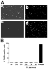
Effect of Lip-C6 on HCE cell apoptosis. (A) HCE cells were incubated with DNase I (positive control; a) or with 5 μM Lip-C6 (b), and apoptosis was detected by TUNEL assay. (c and d) Corresponding DAPI-stained cultures, which identify cell nuclei. (B) Two hundred cells were counted, and the percent TUNEL-positive cells is shown. Note that Lip-C6 did not induce apoptosis in these cells.
Fig. 5
Fig. 5
The effect of Lip-C6 on S. aureus- and LPS-induced inflammation on human and murine neutrophils. (A and B) The human neutrophil cell line (HL-60) was preincubated with 4, 8, or 16 μM Lip-C6 or ghost liposomes 30 min prior to stimulation with S. aureus (A) or LPS (B). After 6 h, CXCL8/IL-8, in cell-free supernatants, was measured by ELISA. Results are mean ± SD of three replicate wells, and data are representative of three independent experiments. Note that Lip-C6 inhibited CXC chemokine production by TLR2- and TLR4-stimulated human neutrophils compared with ghost liposomes. (C–E) Murine peritoneal neutrophils (>95% pure) were incubated overnight with S. aureus (C and D) or with LPS (E and F) in the presence of Lip-C6 or ghost liposomes. CXCL1/keratinocyte-derived chemokine (KC; C and E) and CXCL2/MIP-2 (D and F) were measured by ELISA. Results are mean ± SD of three replicate wells per sample, and data are representative of three independent experiments. P values show significant differences between ghost and Lip-C6 groups at 15 μM.
Fig. 6
Fig. 6
The effect of subconjunctival and topical application of Lip-C6 on neutrophil recruitment to the corneal stroma in mouse models of S. aureus- and LPS-induced corneal inflammation. Lip-C6 (2 nMoles; 811 ng) or ghost liposomes were injected into the subconjunctival space, topically applied to a 1-mm diameter region in the central cornea, or were applied by both methods. After 40 min, corneas were stimulated with inactivated S. aureus (upper panels) or LPS (lower panels), and all were given a second topical application 6 h later. After 24 h, eyes were snap-frozen, and neutrophil numbers in a 5-μm corneal section were determined after immunostaining. Data points represent individual corneas from groups of five or six mice. Note that corneas given Lip-C6 by each of these protocols had significantly less neutrophils than control ghost liposomes. The experiment was repeated three times with similar results.
Fig. 7
Fig. 7
Effect of topical Lip-C6 on S. aureus- and LPS-induced, increased corneal thickness and haze. A wound (1 mm diameter) was made in the central corneal stroma of C57BL/6 mice, and 2 nMoles (811 ng) topical Lip-C6 was given 1 h before and 6 h after exposure to S. aureus (upper panels) or LPS (lower panels). After 24 h, neutrophils were detected by immunohistochemistry, corneas were by in vivo confocal microscopy, and corneal thickness and haze were calculated as described in Materials and Methods. Data points represent individual corneas from groups of four to eight mice, and the experiment was repeated twice with similar results.
Fig. 8
Fig. 8
Therapeutic (post-induction) effect of Lip-C6 on S. aureus-induced neutrophil recruitment. Corneal inflammation was initiated by abrasion and exposure to S. aureus as before. Lip-C6 or ghost liposome (Gh) control 2 nMoles (811 ng) was added topically after 6 h, and eyes were processed for immunohistochemistry after 24 h. Data points represent individual corneas from five mice per group, and the experiment was repeated twice with similar results.
Fig. 9
Fig. 9
Effect of Lip-C6 on corneal epithelial wound healing. A 1-mm diameter corneal epithelial abrasion was made, and corneas were treated with 2 nMoles (811 ng) topical Lip-C6 or ghost liposomes 40 min prior to and 1 and 6 h after exposure to S. aureus. A fluorescein solution (0.25%) was added after 0 h, 6 h, or 24 h to detect the epithelial wound, and the diameter was calculated by image analysis. (A) Representative images of fluorescein binding at indicated time-points. (B) Diameter of wound treated with LipC6 or ghost liposomes (mean of five corneas). Note that there is no inhibitory effect of Lip-C6 on the rate of wound healing. (C) Representative H&E-stained corneal sections 6 h and 24 h after epithelial (Epi) abrasion and exposure to Lip-C6. Note the loss of epithelium at 6 h and regeneration after 24 h; no differences in epithelial regeneration were noted among groups treated with Lip-C6 or controls (data no shown). (D) Corneal sections were also stained with TUNEL reagents (green) to determine the effect on epithelial cell apoptosis, and nuclei were stained with DAPI (blue). Representative sections are shown of naïve corneas (a) or corneal sections after treatment with Lip-C6 and S. aureus (b). TUNEL-positive cells were detected in the stroma and not the epithelium of S. aureus-treated mice. These data are representative of three independent experiments.

References

    1. Fuortes M, Jin W, Nathan C. Ceramide selectively inhibits early events in the response of human neutrophils to tumor necrosis factor. J Leukoc Biol. 1996;59:451–460. - PubMed
    1. Suchard SJ, Mansfield PJ, Boxer LA, Shayman JA. Mitogen-activated protein kinase activation during IgG-dependent phagocytosis in human neutrophils: inhibition by ceramide. J Immunol. 1997;158:4961–4967. - PubMed
    1. Nakamura T, Abe A, Balazovich KJ, Wu D, Suchard SJ, Boxer LA, Shayman JA. Ceramide regulates oxidant release in adherent human neutrophils. J Biol Chem. 1994;269:18384–18389. - PubMed
    1. Wong K, Li XB, Hunchuk N. N-acetylsphingosine (C2-ceramide) inhibited neutrophil superoxide formation and calcium influx. J Biol Chem. 1995;270:3056–3062. - PubMed
    1. Sun Y, Hise AG, Kalsow CM, Pearlman E. Staphylococcus aureus-induced corneal inflammation is dependent on Toll-like receptor 2 and myeloid differentiation factor 88. Infect Immun. 2006;74:5325–5332. - PMC - PubMed

Publication types

MeSH terms