. 2008 Jun;22(6):1449-63.
doi: 10.1210/me.2008-0035. Epub 2008 Mar 27.

Protein kinase Calpha-induced derepression of the human luteinizing hormone receptor gene transcription through ERK-mediated release of HDAC1/Sin3A repressor complex from Sp1 sites

Affiliations

Protein kinase Calpha-induced derepression of the human luteinizing hormone receptor gene transcription through ERK-mediated release of HDAC1/Sin3A repressor complex from Sp1 sites

Mingjuan Liao et al. Mol Endocrinol. 2008 Jun.

Abstract

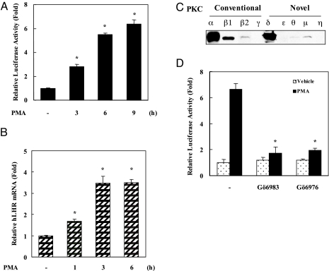
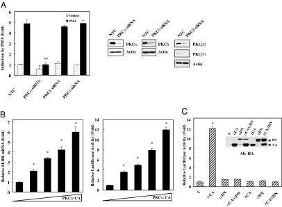
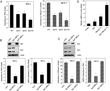
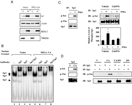
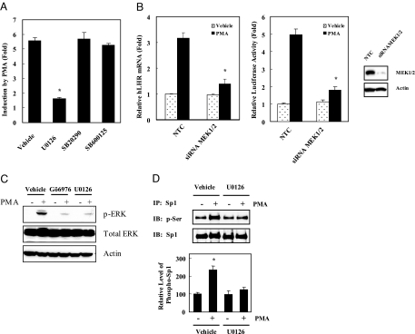
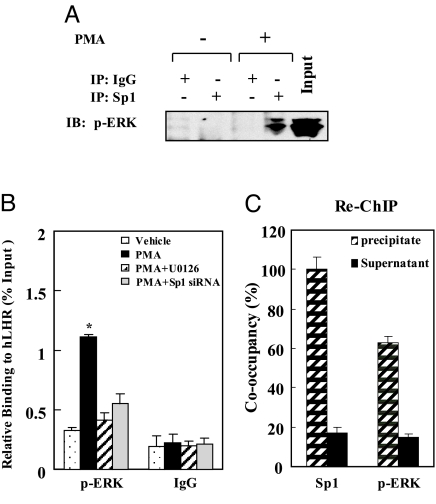
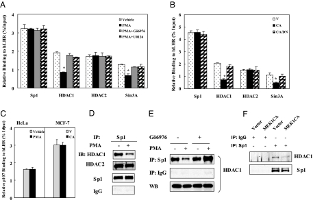
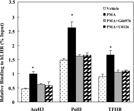
LH receptor (LHR) gene transcription is subject to repression/derepression through various modes and multiple effectors. Epigenetic silencing and activation of the LHR is achieved through coordinated regulation at both histone and DNA levels. The LHR gene is subject to repression by deacetylation and methylation at its promoter region, where a HDAC/mSin3A repressor complex is anchored at Sp1 sites. The present studies revealed that protein kinase C (PKC) alpha/ERK signaling is important for the activation of LHR promoter activity, and the increase of endogenous transcripts induced by phorbol-12-myristate-13-acetate (PMA) in HeLa cells. Whereas these effects were attributable to PKCalpha activity, the ERK pathway was the downstream effector in LHR activation. PMA caused a significant enhancement of Sp1 phosphorylation at serine residue (s), which was blocked by PKCalpha or ERK inhibition. The interaction of activated phosphorylated ERK with Sp1 and ERK's association with the LHR promoter points to Sp1 as a direct target of ERK. After Sp1 phosphorylation, the HDAC1/mSin3A repressor complex dissociated from Sp1 sites, histone 3 was acetylated, and transcription factor II B and RNA polymerase II were recruited. In addition, overexpression of a constitutively active PKCalpha (PKCalpha CA) strongly activated LHR transcription in MCF-7 cells (devoid of PKCalpha), induced Sp1 phosphorylation at serine residue (s) and caused derecruitment of HDAC1/mSin3A complex from the promoter. These effects were negated by cotransfection of a dominant-negative PKCalpha. In conclusion, these studies have revealed a novel regulatory signaling mechanism of transcriptional control in which the LHR is derepressed through PKCalpha/ERK-mediated Sp1 phosphorylation, causing the release of HDAC1/mSin3A complex from the promoter.


Figures

Figure 1
Figure 1
Effect of PKC Activation by PMA on Transcription of the hLHR Gene A, Reporter gene analyses of hLHR gene promoter activity in response to PMA in HeLa cells. At 36 h after transfection with the wild-type hLHR promoter/reporter gene construct, cells were incubated with 100 nm PMA for 0–9 h. Relative luciferase activity was expressed as fold-induction (n-fold) over the activity in the absence of PMA (1-fold). Results shown in this and subsequent figures are the mean ± se of three independent experiments in triplicate (*, P < 0.05). B, Real-time PCR analyses of hLHR expression in HeLa cells treated with 100 nm PMA or vehicle for 0–6 h. The relative mRNA levels are shown as n-fold induction over the levels in the absence of treatment (1-fold) (*, P < 0.05). C, Western blot analyses of endogenous expression of PKC isoforms. D, Reporter gene analyses in HeLa cells pretreated with or without PKC inhibitors (Gö6983, Gö6976) for 1 h before incubation with PMA (100 nm) or vehicle for additional 9 h. Relative luciferase activity was expressed as fold-induction over the activity in the absence of treatment (*, P < 0.05, compared with the PMA-induced activity in the absence of inhibitor).
Figure 2
Figure 2
PKCα Is Required for PMA Activation of the hLHR Gene A, Reporter gene analysis of LHR gene promoter activity in HeLa cells cotransfected with PKCα, PKCβ, PKCδ siRNA, or negative control siRNA (NTC) incubated with and without PMA (100 nm) for 9 h. Relative luciferase activity was expressed as fold-induction over the promoter activity with vehicle treatment. Expression levels of PKCα, PKCβ1/2, and PKCδ in HeLa cells transfected with siRNA of PKCα, PKCβ, PKCδ, and NTC were analyzed by Western blot analyses (right) (*, P < 0.05, compared with NTC without PMA treatment. **, P < 0.05, compared with NTC with PMA treatment). B, Real-time PCR analyses of hLHR gene expression (left) and reporter gene analyses of its promoter activity (right) in MCF-7 cells that were cotransfected with increasing amount of PKCα CA (*, P < 0.05). C, Reporter gene analyses of hLHR promoter activity in MCF-7 cells that were transfected with CA or DN PKC constructs alone or both. HA-tag expression of transfected PKCα or PKCδ constructs was also shown. V, Empty vector; Ab, antibody; HA, hemagglutinin. (αCA, PKCαCA; δCA, PKCδCA; αDN, PKCαDN; δDN, PKCδ DN) (*, P < 0.05).
Figure 3
Figure 3
Requirement of Both Sp1/Sp3 Binding Sites and Involvement of Sp1 But Not Sp3 in PMA-Mediated hLHR Promoter Activation A, Participation of Sp1/Sp3 binding sites Sp1(I) and Sp1(II) on the activation of hLHR promoter activity by PMA in HeLa cells (left) and by PKCα CA in MCF-7 cells (right). Reporter gene analyses of HeLa cells transfected with the wild-type hLHR promoter construct (WT) or with mutations on either Sp1(I) site (Sp1X) or Sp1(II) site (Sp2X) or on both sites (Sp1–2X), treated with vehicle or 100 nm PMA for 9 h. In MCF-7 cells, the hLHR promoter WT or mutated constructs were cotransfected with empty pcDNA 3.1 vector or PKCα CA. Luciferase activity was determined 36 h after transfection. Data were shown as the fold-induction of luciferase activity by PMA treatment (HeLa cells) or PKCα CA overexpression (MCF-7 cells) with the different promoter constructs over control in the absence of PMA (HeLa) or empty vector (MCF-7) (*, P < 0.05). B and C, Reporter gene analyses of hLHR promoter activity (left) and real-time PCR analyses of its expression (right) in HeLa and MCF-7 cells cotransfected with Sp1, Sp3 siRNA or NTC. HeLa cells were treated with 100 nm PMA or vehicle for 9 h. MCF-7 cells were cotransfected with pcDNA3.1 empty vector or PKCα CA. Relative luciferase activity was expressed as fold-induction over the promoter activity in the absence of PMA in HeLa cells or without PKCα CA in MCF-7 cells. Expression of Sp1 and Sp3 proteins in HeLa cells (B, top) and MCF-7 cells (C, top) transfected with Sp1, Sp3 or NTC siRNA was analyzed by Western blot analyses. The expression levels of β-actin served as loading control. (*, P < 0.05). D, Synergistic effect of Sp1 on PKCα CA-induced hLHR activation. Quantitative real-time PCR analyses of hLHR mRNA from MCF-7 cells transfected with empty vector only, Sp1 or pKCα CA or both. The relative amount of hLHR mRNA was expressed as fold-increase over the mRNA amount with transfection of pcDNA 3.1 empty vector (*, P < 0.05).
Figure 4
Figure 4
PMA Treatement Enhances Sp1 Phosphorylation in a PKCα-Dependent Manner A, Western blot analyses of cytosolic and nuclear extracts from MCF-7 cells overexpressing PKCα CA or vector using Sp1, Sp3, and β-actin antibodies. Expression of HDAC1 and tubulin, as nuclear and cytoplasmic protein markers, respectively, are also shown. B, EMSA of Sp1/Sp3 binding to the Sp1(I) site of the hLHR promoter. 32P-labeled double-strand DNA oligomer containing the Sp1(I) site of the hLHR promoter was incubated with nuclear extracts prepared from MCF-7 cells transfected with empty pcDNA 3.1 (vector) only, or pKCα CA. The reaction was carried out in the absence or presence of antibodies to Sp1, Sp3 or both. C, Effects of PMA on the phosphorylation of Sp1 in HeLa cells. Left, Nuclear extracts from PMA-treated (100 nm, 3 h) (+) or untreated (−) HeLa cells were immunoprecipitated (IP) with Sp1 antibody. Samples (immunoprecipitates) were analyzed by Western blots (IB) using p-Ser, p-Thr, and Sp1 antibodies. Right, Nuclear extracts from HeLa cells stimulated with 100 nm PMA (+) or vehicle (−) for 3 h in the presence or absence of Gö6976 were immunoprecipitated with Sp1 antibody. The immunocomplex was analyzed by Western blots for detection of Sp1 and p-Sp1. The relative levels of p-Sp1 were quantified by densitometry (*, P < 0.05). D, Effects of PKCα CA overexpression in MCF-7 cells on the phosphorylation of Sp1. Left, Nuclear extracts from MCF-7 cells that were transfected with empty vector (V) or PKCα CA were immunoprecipitated with Sp1 antibody, followed by Western blots using p-Ser, p-Thr, and Sp1 antibodies. Right, Nuclear extracts from MCF-7 cells overexpressing PKCα CA in the presence or absence of PKCα DN or vector only were immunoprecipitated by Sp1 antibody or normal IgG. The immunocomplexes were analyzed by Western blots using Sp1 and p-Ser antibodies.
Figure 5
Figure 5
ERK/MAPK Pathway Participates in the PMA-Induced hLHR Activation A, Reporter gene analyses of HeLa cells preincubated with or without 10 μm of inhibitors to ERK (U0126), p38 (SB20290), and JNK (SB600125) for 1 h before simulation with 100 nm PMA for 9 h (*, P < 0.05). B, Quantitative real-time PCR analyses of hLHR gene expression (left) and reporter gene analyses of its promoter activity (middle) in HeLa cells transfected NTC or MAPK kinase (MEK) 1/2 siRNA. Cells were treated with 100 nm PMA or vehicle for 9 h. Expression of MEK1/2 in HeLa cells transfected with siRNA of MEK1/2 or NTC analyzed by Western blot (right). (*, P < 0.05, compared with NCT treated with PMA). C, HeLa cells were pretreated with or without Gö6976 or U0126 for 1 h, followed by incubation with 100 nm PMA or vehicle for 15 min. Cell lysates were analyzed by Western blot using antibodies against p-ERK1/2, total ERK, and β-actin. D, Immunoprecipitation (IP) of Sp1 in nuclear extracts from HeLa cells treated with 100 nm PMA (+) or vehicle (−) for 3 h in the presence or absence of U0126. The phosphorylation level of Sp1 in the immunocomplex was determined by Western blot (IB) analyses with anti-p-Ser antibody. The total Sp1 immunoprecipitated is also shown. The relative level of p-Sp1 was quantified by densitometry (*, P < 0.05).
Figure 6
Figure 6
ERK Interacts with Sp1 and Is Recruited to the hLHR Promoter in the Presence of PMA A, Interaction of p-ERK and Sp1 determined by immunoprecipitation (IP). Whole cell lysates prepared from HeLa cells treated with PMA (+) or vehicle (−) for 15 min were immunoprecipitated with normal IgG or Sp1 antibody. The immunocomplex was analyzed by Western blot (IB) analyses with p-ERK antibody. The expression of p-ERK in the cells stimulated with PMA for 15 min is shown as input. B, Association of p-ERK with hLHR promoter by ChIP analyses. HeLa cells were treated with 100 nm PMA or vehicle for 15 min in the presence and absence of MEK inhibitor U0126 (10 μm), or Sp1 siRNA, and subjected to chromatin immunoprecipitation analyses with p-ERK antibody or normal rabbit IgG. The precipitated DNA was analyzed quantitatively by real-time PCR with primers amplifying the core promoter sequence of hLHR gene. The relative binding to promoter was expressed as the percentage amount over input (%). Data were presented as the mean ± se from three independent experiments in triplicate (*, P < 0.05). C, Association of p-ERK to Sp1 bound to the LHR promoter determined Re-ChIP analyses. Sequential ChIP analyses of HeLa cells treated by PMA for 15 min using Sp1 as the first antibody followed by the second immunoprecipitation with p-ERK antibody. The DNA precipitates and supernatants after the second immunoprecipitation were examined by quantitative PCR, respectively. Co-occupancy of Sp1 set to 100% is also shown.
Figure 7
Figure 7
PMA Treatment Leads to PKCα/ERK-Dependent Release of HDAC1/mSin3A Complex from the hLHR Promoter A and B, Quantitative ChIP analyses in HeLa cells (A) and MCF-7 cells (B). HeLa cells were treated with 100 nm PMA or vehicle for 3 h in the presence or absence of Gö6976 or U0126. MCF-7 cells were transfected with pcDNA3.1 vector (V) or PKCα CA (CA) alone or cotransfected with PKCα DN (CA/DN). Cells were subjected to ChIP assay with various antibodies (Sp1, HDAC1, HDAC2, and mSin3A). The precipitated DNA was analyzed by real-time PCR with primers for hLHR gene promoter. The relative binding of these factors with the promoter was presented as the percentage amount over input (%) (*, P < 0.05). C, Quantitative ChIP analyses of p107 association to hLHR promoter in HeLa cells treated with vehicle or PMA (100 nm, 3 h) (left) and MCF-7 cells transfected with pcDNA 3.1 vector (V) or PKCα CA (CA) (right). D, Nuclear extracts were prepared from HeLa cells treated with vehicle (−) or 100 nm PMA (+) for 3 h, and immunoprecipitated with Sp1 antibody. The immunocomplex was analyzed by Western blot analyses with HDAC1, HDAC2 or Sp1 antibodies. E, HeLa cells were pretreated with or without Gö6976 for 1 h before exposure to vehicle (−) or 100 nm PMA (+) for 3 h. Nuclear extracts were immmunoprecipitated with Sp1 antibody or IgG followed by Western blot analyses with HDAC1 antibody. Expression level of HDAC1 in these cells was shown as input. F, Nuclear extracts from MCF-7 cells transfected with vector or MEK1CA were immunoprecipitated with Sp1 antibody followed by Western blot analyses with HDAC1 antibody. Total immunoprecipitated Sp1 level was also shown.
Figure 8
Figure 8
PMA Treatment Increases Acetylation of Histone H3, and Recruitment of Pol II and TFIIB to the LHR Promoter HeLa cells were treated with vehicle or 100 nm PMA for 3 h in the presence or absence of Gö6976 (2 μm) or U0126 (10 μm), and subjected to quantitative ChIP analyses of AceH3, Pol II, and TFIIB at the hLHR promoter. The relative binding of indicated transcriptional factors was expressed as the percentage amount over input (%). Data were presented as the mean ± se from three independent experiments in triplicate (*, P < 0.05).

Similar articles

Cited by

References

    1. Dufau ML, Tsai-Morris C-H 2007 The Leydig cell in health and disease. In: Payne AH, Hardy MP, eds. Contemporary endocrinology. Totowa, NJ: Humana Press Inc.; 227–252
    1. Dufau ML 1998 The luteinizing hormone receptor. Annu Rev Physiol 60:461–496 - PubMed
    1. Tsai-Morris C, Xie X, Wang W, Buczko E, Dufau M 1993 Promoter and regulatory regions of the rat luteinizing hormone receptor gene. J Biol Chem 268:4447–4452 - PubMed
    1. Tsai-Morris C, Geng Y, Xie X, Buczko E, Dufau M 1994 Transcriptional protein binding domains governing basal expression of the rat luteinizing hormone receptor gene. J Biol Chem 269:15868–15875 - PubMed
    1. Tsai-Morris CH, Geng Y, Buczko E, Dufau ML 1998 A novel human luteinizing hormone receptor gene. J Clin Endocrinol Metab 83:288–293 - PubMed

Publication types

MeSH terms