

Case Reports
doi: 10.3174/ajnr.A1074.
Epub 2008 Mar 27.
A developmental venous anomaly presenting atypical findings on susceptibility-weighted imaging
- PMID: 18372413
- PMCID: PMC8119127
- DOI: 10.3174/ajnr.A1074
Item in Clipboard
Case Reports
A developmental venous anomaly presenting atypical findings on susceptibility-weighted imaging
AJNR Am J Neuroradiol.
2008 Aug.
No abstract available
Figures
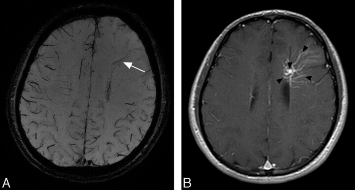
A 26-year-old man with a DVA. A, MIP image of SWI shows a moderate low-signal-intensity structure corresponding to the draining vein (arrow); however, no low-signal-intensity structures corresponding to medullary veins are seen. Low-signal-intensity structures corresponding to the cortical veins of the other lobes are clearly shown; however, no apparent signals of cortical veins in the left frontal lobe are identified. B, Postcontrast T1-weighted images at 1.5T obtained 2 days after precontrast images reveal the enhanced draining vein (arrow) and medullary veins (arrowheads).
Comment in
-
When is an atypical DVA an AVM?AJNR Am J Neuroradiol. 2009 Feb;30(2):E24; author reply E25. doi: 10.3174/ajnr.A1310. Epub 2008 Nov 27. AJNR Am J Neuroradiol. 2009. PMID: 19039038 Free PMC article. No abstract available.
References
Publication types
MeSH terms
Substances
LinkOut - more resources
Full Text Sources
Medical