

Subtraction 3D CT angiography with the orbital synchronized helical scan technique for the evaluation of postoperative cerebral aneurysms treated with cobalt-alloy clips
- PMID: 18372416
- PMCID: PMC8118823
- DOI: 10.3174/ajnr.A1040
Subtraction 3D CT angiography with the orbital synchronized helical scan technique for the evaluation of postoperative cerebral aneurysms treated with cobalt-alloy clips
Abstract
Background and purpose: CT angiography (CTA) has been used for the evaluation of intracranial aneurysms and recently has been applied to assess postoperative aneurysms treated with titanium-alloy clips. We investigated the clinical usefulness of subtraction CTA by using the orbital synchronized helical scan technique (OSHST) for evaluating intracranial aneurysms surgically treated with cobalt-alloy clips.
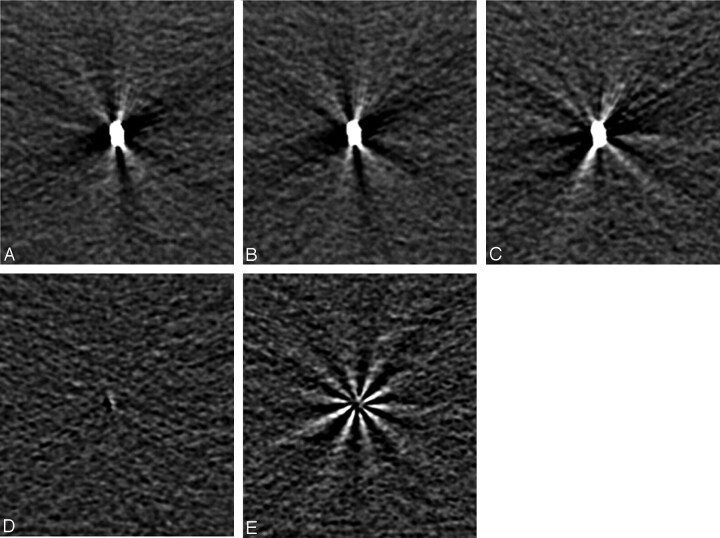
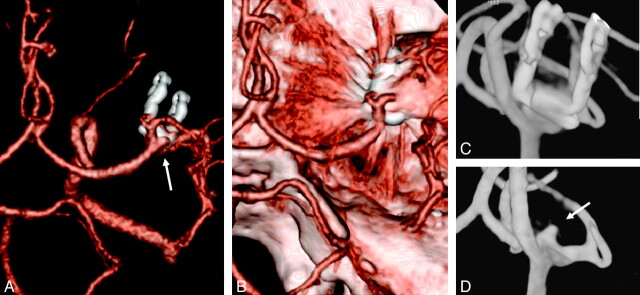
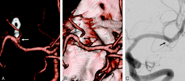
Materials and methods: We scanned an agar gel phantom with a cobalt-alloy clip mounted in the center by using subtraction CT with and without OSHST. Eighteen patients (20 aneurysms) who underwent surgery with cobalt-alloy clips were postoperatively evaluated with subtraction CTA with OSHST, and the results were compared with those from digital subtraction angiography. Two neuroradiologists independently evaluated the 3D CTA images and source images with and without subtraction for the presence of residual flow in the aneurysm and stenotic change in parent or neighboring arteries.
Results: For the phantom study, significantly fewer artifacts from clips were noted on images obtained by using subtraction CT with OSHST than on those obtained without OSHST. For the clinical study, subtraction CTA with OSHST also showed fewer clip artifacts than did conventional CTA. Image quality was poor, and we were unable to diagnose residual neck for 5% (1/20) with subtraction CTA with OSHST and 75% (15/20) with conventional CTA. For evaluation of adjacent vessels, image quality was poor for none (0/20) with subtraction CTA with OSHST and for 55% (11/20) with conventional CTA. For subtraction CTA with OSHST, sensitivity in detecting residual neck was 1.0, and specificity was 0.94. For conventional CTA, sensitivity and specificity were both 0.25.
Conclusions: OSHST is a useful technique for subtracting cobalt-alloy clips, and subtraction CTA with OSHST is available for evaluating aneurysms after clipping with cobalt-alloy clips.
Figures
Similar articles
-
Dual-energy CT angiography in the evaluation of intracranial aneurysms: image quality, radiation dose, and comparison with 3D rotational digital subtraction angiography.AJR Am J Roentgenol. 2010 Jan;194(1):23-30. doi: 10.2214/AJR.08.2290. AJR Am J Roentgenol. 2010. PMID: 20028901
-
The Utility of Dual-Energy Computed Tomographic Angiography for the Evaluation of Brain Aneurysms After Surgical Clipping: A Prospective Study.World Neurosurg. 2015 Nov;84(5):1362-71. doi: 10.1016/j.wneu.2015.06.027. Epub 2015 Jun 23. World Neurosurg. 2015. PMID: 26115801
-
Limitations of three-dimensional reconstructed computerized tomography angiography after clip placement for intracranial aneurysms.J Neurosurg. 2005 Oct;103(4):656-61. doi: 10.3171/jns.2005.103.4.0656. J Neurosurg. 2005. PMID: 16266048
-
Image quality and artefact generation post-cerebral aneurysm clipping using a 64-row multislice computer tomography angiography (MSCTA) technology: A retrospective study and review of the literature.Clin Neurol Neurosurg. 2010 Jun;112(5):386-91. doi: 10.1016/j.clineuro.2010.02.001. Epub 2010 Mar 1. Clin Neurol Neurosurg. 2010. PMID: 20189713 Review.
-
Virtues and drawbacks of titanium alloy aneurysm clips.Acta Neurochir Suppl. 1999;72:81-8. doi: 10.1007/978-3-7091-6377-1_7. Acta Neurochir Suppl. 1999. PMID: 10337415 Review.
Cited by
-
Reduction of misregistration on cerebral four-dimensional computed tomography angiography images using advanced patient motion correction reconstruction.Jpn J Radiol. 2016 Sep;34(9):605-10. doi: 10.1007/s11604-016-0563-1. Epub 2016 Jul 5. Jpn J Radiol. 2016. PMID: 27379502
-
Subtraction CT angiography for the diagnosis of iliac arterial steno-occlusive disease.Jpn J Radiol. 2016 Mar;34(3):194-202. doi: 10.1007/s11604-015-0508-0. Epub 2015 Dec 18. Jpn J Radiol. 2016. PMID: 26682737
-
Metallic Component Preserving Algorithm Based on the Cerebral Computed Tomography Angiography in Aneurysm Surgery.Diagnostics (Basel). 2022 Jan 28;12(2):338. doi: 10.3390/diagnostics12020338. Diagnostics (Basel). 2022. PMID: 35204429 Free PMC article.
-
Cerebral bone subtraction CT angiography using 80 kVp and sinogram-affirmed iterative reconstruction: contrast medium and radiation dose reduction with improvement of image quality.Neuroradiology. 2017 Feb;59(2):127-134. doi: 10.1007/s00234-016-1776-9. Epub 2017 Jan 3. Neuroradiology. 2017. PMID: 28050639
-
3D rotational fluoroscopy for intraoperative clip control in patients with intracranial aneurysms--assessment of feasibility and image quality.BMC Med Imaging. 2016 Apr 19;16:30. doi: 10.1186/s12880-016-0133-0. BMC Med Imaging. 2016. PMID: 27094510 Free PMC article.
References
-
- Venema HW, Hulsmans FJ, den Heeten GJ. CT angiography of the circle of Willis and intracranial internal carotid arteries: maximum intensity projection with matched mask bone elimination-feasibility study. Radiology 2001;218:893–98 - PubMed
-
- Dehdashti AR, Binaghi S, Uske A, et al. Comparison of multislice computerized tomography angiography and digital subtraction angiography in the postoperative evaluation of patients with clipped aneurysms. J Neurosurg 2006;104:395–403 - PubMed
MeSH terms
Substances
LinkOut - more resources
Full Text Sources
Medical