

Retinal dysfunction and progressive retinal cell death in SOD1-deficient mice
- PMID: 18372426
- PMCID: PMC2329841
- DOI: 10.2353/ajpath.2008.070730
Retinal dysfunction and progressive retinal cell death in SOD1-deficient mice
Abstract
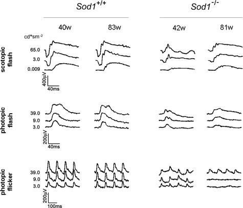
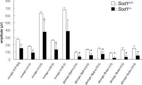
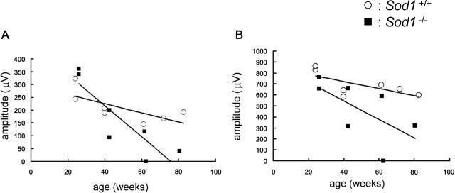
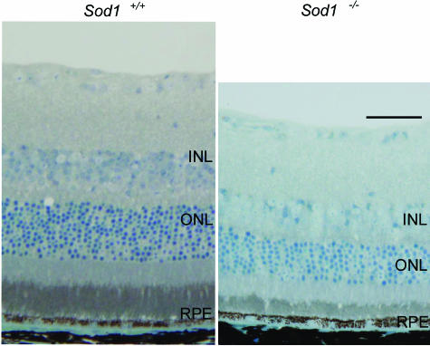
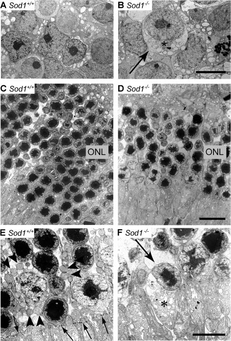
The superoxide dismutase (SOD) family is a major antioxidant system, and deficiency of Cu,Zn-superoxide dismutase (SOD1) in mice leads to many different phenotypes that resemble accelerated aging. The purpose of this study was to examine the morphology and physiology of the sensory retina in Sod1(-/-) mice. The amplitudes of the a- and b-waves of electroretinograms elicited by stimuli of different intensity were reduced in senescent Sod1(-/-) mice, and this reduction in amplitude was more pronounced with increasing age. Retinal morphometric analyses showed a reduced number of nuclei in both the inner nuclear cell layer and outer nuclear cell layer. Electron microscopy revealed swollen cells and degenerated mitochondria in the inner nuclear cell and outer nuclear cell layer of senescent Sod1(-/-) mice indicating necrotic cell death. Terminal deoxynucleotidyl transferase-mediated dUTP nick end labeling revealed no significant differences in the number of apoptotic cells between Sod1(-/-) and wild-type mice, and activated caspase-3 could not be detected in the retina of Sod1(-/-) mice. In addition to the age-related macular degeneration-like phenotypes previously reported, Sod1(-/-) mice also present progressive retinal degeneration. Our results indicate that Sod1(-/-) mice may be a good model system in which to study the mechanism of reactive oxygen species-mediated retinal degeneration.
Figures
Similar articles
-
Neuroprotective role of superoxide dismutase 1 in retinal ganglion cells and inner nuclear layer cells against N-methyl-d-aspartate-induced cytotoxicity.Exp Eye Res. 2013 Oct;115:230-8. doi: 10.1016/j.exer.2013.07.002. Epub 2013 Jul 13. Exp Eye Res. 2013. PMID: 23856406
-
Mice With a Combined Deficiency of Superoxide Dismutase 1 (Sod1), DJ-1 (Park7), and Parkin (Prkn) Develop Spontaneous Retinal Degeneration With Aging.Invest Ophthalmol Vis Sci. 2019 Sep 3;60(12):3740-3751. doi: 10.1167/iovs.19-27212. Invest Ophthalmol Vis Sci. 2019. PMID: 31487745 Free PMC article.
-
Age-related dysfunction of the lacrimal gland and oxidative stress: evidence from the Cu,Zn-superoxide dismutase-1 (Sod1) knockout mice.Am J Pathol. 2012 May;180(5):1879-96. doi: 10.1016/j.ajpath.2012.01.019. Epub 2012 Mar 20. Am J Pathol. 2012. PMID: 22440255
-
Superoxide dismutase 1 protects retinal cells from oxidative damage.J Cell Physiol. 2006 Sep;208(3):516-26. doi: 10.1002/jcp.20683. J Cell Physiol. 2006. PMID: 16741961
-
Vision deficits precede structural losses in a mouse model of mitochondrial dysfunction and progressive retinal degeneration.Exp Eye Res. 2011 Dec;93(6):833-41. doi: 10.1016/j.exer.2011.09.017. Epub 2011 Oct 5. Exp Eye Res. 2011. PMID: 21983042
Cited by
-
Glutathione peroxidase 4 is required for maturation of photoreceptor cells.J Biol Chem. 2012 Mar 2;287(10):7675-82. doi: 10.1074/jbc.M111.335174. Epub 2011 Dec 29. J Biol Chem. 2012. PMID: 22207760 Free PMC article.
-
Expression of 8-oxoguanine DNA glycosylase (Ogg1) in mouse retina.Mol Vis. 2009 Jun 5;15:1139-52. Mol Vis. 2009. PMID: 19503746 Free PMC article.
-
The M1 muscarinic acetylcholine receptor subtype is important for retinal neuron survival in aging mice.Sci Rep. 2019 Mar 26;9(1):5222. doi: 10.1038/s41598-019-41425-5. Sci Rep. 2019. PMID: 30914695 Free PMC article.
-
Aging related changes of retina and optic nerve of Uromastyx aegyptia and Falco tinnunculus.ACS Chem Neurosci. 2014 Jan 15;5(1):39-50. doi: 10.1021/cn400154k. Epub 2013 Nov 19. ACS Chem Neurosci. 2014. PMID: 24215233 Free PMC article.
-
Unraveling a multifactorial late-onset disease: from genetic susceptibility to disease mechanisms for age-related macular degeneration.Annu Rev Genomics Hum Genet. 2009;10:19-43. doi: 10.1146/annurev.genom.9.081307.164350. Annu Rev Genomics Hum Genet. 2009. PMID: 19405847 Free PMC article. Review.
References
-
- Valentine JS, Doucette PA, Potter SZ. Copper-zinc superoxide dismutase and amyotrophic lateral sclerosis. Annu Rev Biochem. 2005;74:563–593. - PubMed
-
- Behndig A, Svensson B, Marklund SL, Karlsson K. Superoxide dismutase isoenzymes in the human eye. Invest Ophthalmol Vis Sci. 1998;39:471–475. - PubMed
-
- Chang B, Hawes NL, Hurd RE, Davisson MT, Nusinowitz S, Heckenlively JR. Retinal degeneration mutants in the mouse. Vision Res. 2002;42:517–525. - PubMed
-
- Dalke C, Graw J. Mouse mutants as models for congenital retinal disorders. Exp Eye Res. 2005;81:503–512. - PubMed
-
- Grooten JV, Goossens B, Vanhaesebroeck B, Fiers W. Cell membrane permeabilization and cellular collapse, followed by loss of dehydrogenase activity: early events in tumor necrosis factor-induced cytotoxicity. Cytokine. 1993;5:546–555. - PubMed
MeSH terms
Substances
LinkOut - more resources
Full Text Sources
Molecular Biology Databases
Research Materials
Miscellaneous
