Comparative Study
. 2008 May;40(5):600-8.
doi: 10.1038/ng.115. Epub 2008 Mar 30.

Differential effects of oncogenic K-Ras and N-Ras on proliferation, differentiation and tumor progression in the colon

Affiliations
Comparative Study

Differential effects of oncogenic K-Ras and N-Ras on proliferation, differentiation and tumor progression in the colon

Kevin M Haigis et al. Nat Genet. 2008 May.

Abstract

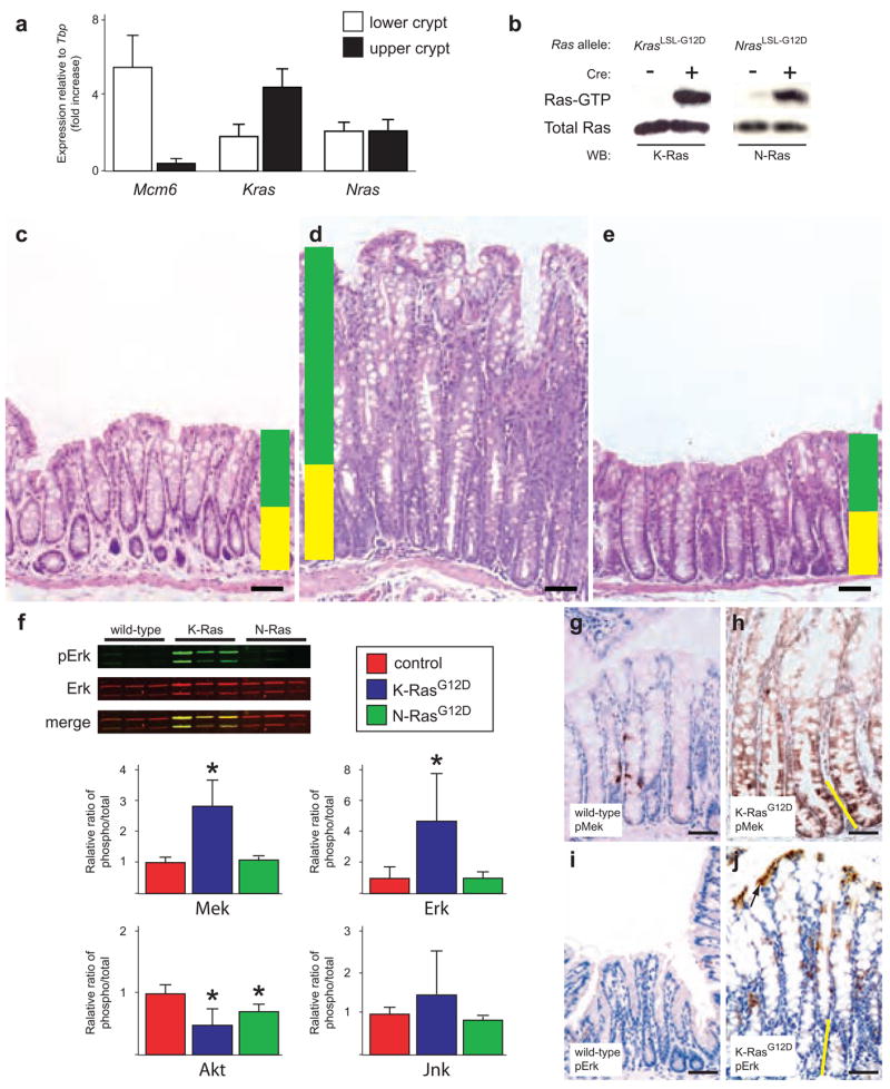
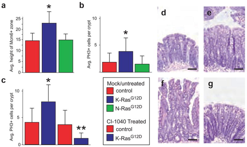
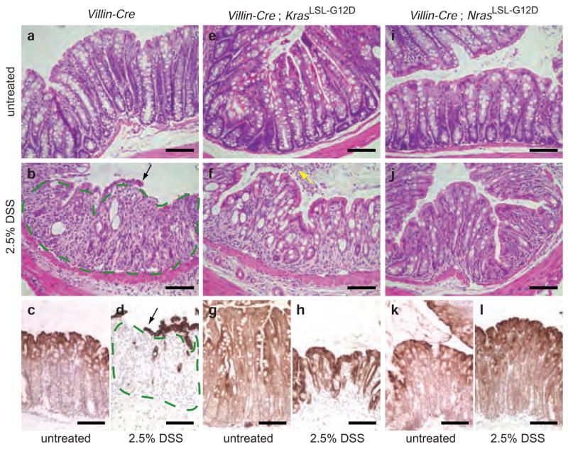
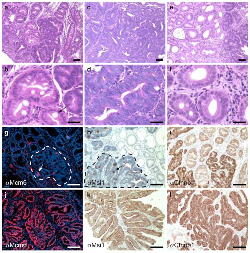
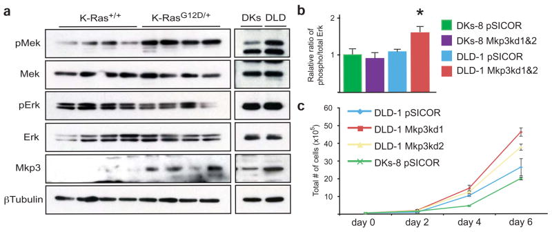
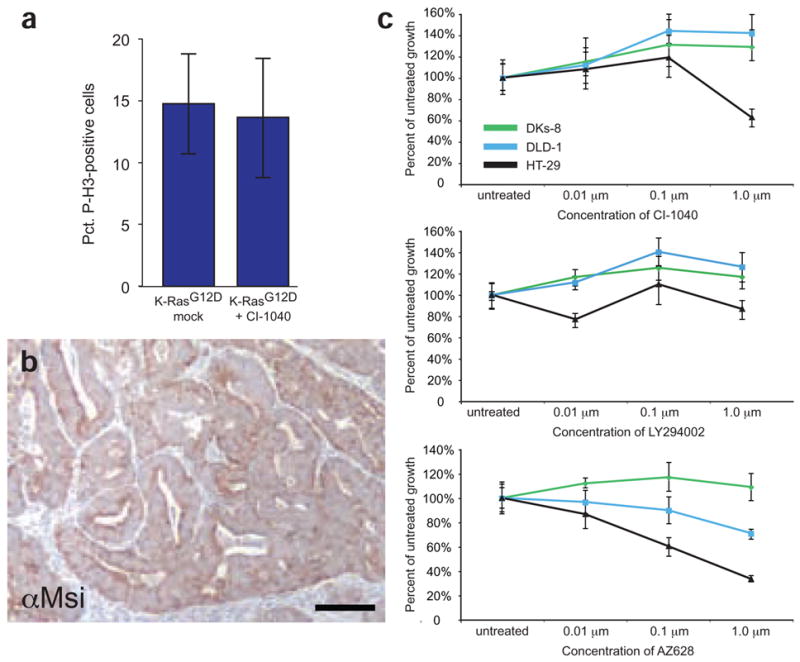
Kras is commonly mutated in colon cancers, but mutations in Nras are rare. We have used genetically engineered mice to determine whether and how these related oncogenes regulate homeostasis and tumorigenesis in the colon. Expression of K-Ras(G12D) in the colonic epithelium stimulated hyperproliferation in a Mek-dependent manner. N-Ras(G12D) did not alter the growth properties of the epithelium, but was able to confer resistance to apoptosis. In the context of an Apc-mutant colonic tumor, activation of K-Ras led to defects in terminal differentiation and expansion of putative stem cells within the tumor epithelium. This K-Ras tumor phenotype was associated with attenuated signaling through the MAPK pathway, and human colon cancer cells expressing mutant K-Ras were hypersensitive to inhibition of Raf, but not Mek. These studies demonstrate clear phenotypic differences between mutant Kras and Nras, and suggest that the oncogenic phenotype of mutant K-Ras might be mediated by noncanonical signaling through Ras effector pathways.


Figures

Figure 1
Figure 1
Mutant K-Ras, but not mutant N-Ras, promotes hyperplastic growth in the colonic epithelium. (a) Expression analysis of Kras and Nras in the colonic epithelium. We used Mcm6, which is expressed only in undifferentiated cells at the bottom of the crypt, as a control. Kras is more highly expressed at the top of the crypt, whereas Nras is uniformly expressed throughout the crypt. Expression of Mcm6, Kras and Nras was normalized to the expression of the TATA-box binding protein (Tbp) gene. (b) Biochemical detection of activated K-Ras and N-Ras. Activated (GTP-bound) forms of each protein can be detected in lysates from colonic epithelium, but only in mice carrying the conditional (LSL-G12D) allele and expressing Cre recombinase from the Fabpl promoter. (c) Hematoxylin and eosin staining of normal colonic epithelium. Yellow shading denotes the normal proliferative zone, and green shading marks the zone of differentiation. All animals were killed sacrificed between 4–6 months of age. (d) Hematoxylin and eosin staining of colonic epithelium expressing activated K-RasG12D showing crypt hyperplasia. (e) Hematoxylin and eosin staining of colonic epithelium expressing activated N-RasG12D. (f) Signaling downstream of K-RasG12D and N-RasG12D in vivo. In quantitative protein blots of whole tissue lysates (see example in upper left), mutant K-Ras activates Mek and Erk. Mutant N-Ras, by contrast, does not activate Mek or Erk. Both K-RasG12D and N-RasG12D downregulate phospho-Akt, whereas neither seems to significantly affect phospho-Jnk. *P < 0.05, Wilcoxon rank sum test. The activation state of each molecule is measured as the ratio of phospho-protein to total protein. (g,h) Immunohistochemical detection of phospho-Mek. Few cells in the normal colonic epithelium (g) express phospho-Mek, whereas all cells expressing mutant K-Ras stain positively (h). (i,j) Immunohistochemical detection of phospho-Erk. Phospho-Erk is readily detectable in the differentiated cells at the top of the colonic crypt expressing K-RasG12D (j), but not in the proliferative cells at the bottom of the crypt. In h and j, the height of the proliferative progenitor zone is represented by the yellow line. Scale bar in all panels, 50 μm. Error bars in panels a and f represent standard deviations for each sample class.
Figure 2
Figure 2
K-RasG12D promotes hyperproliferation through Mek. (a) Quantification of the number of Mcm6-positive cells at the bottom of the crypt shows that mutant K-Ras, but not mutant N-Ras, significantly increases the size of the progenitor cell population. *P < 0.01, Wilcoxon rank sum test. (b) The progenitor cells expressing K-RasG12D are hyperproliferative, as assessed by the number of cells per crypt that are positive for phosphorylated histone-H3, a marker of mitotic cells. * P < 0.01. (c) Inhibition of Mek with CI-1040 suppresses proliferation in colonic epithelium expressing mutant K-Ras. **P < 10−24, Wilcoxon rank sum test. Inhibition of Mek has no effect on proliferation in control colons. (d) Hematoxylin and eosin staining of normal, untreated colonic epithelium. (e) Hematoxylin and eosin staining of wild-type colon in an animal treated with CI-1040 for 24 h. (f) Hematoxylin and eosin staining of colonic epithelium expressing activated K-RasG12D showing crypt hyperplasia. (g) Hematoxylin and eosin staining of K-RasG12D colonic epithelium treated with CI-1040 for 24 h. Note the absence of hyperplasia and the return to somewhat normal histology. Scale bar in all panels, 50 μm. Error bars in panels a-c represent standard deviations for each sample class.
Figure 3
Figure 3
Activated N-Ras, but not K-Ras, suppresses DSS-induced apoptosis in the colonic epithelium. (a) Hematoxylin and eosin staining of colonic epithelium from an untreated Villin-Cre mouse. Villin-Cre directs expression of Cre throughout the entire small intestinal and colonic epithelia. (b) Hematoxylin and eosin staining of colonic epithelium from a Villin-Cre mouse treated with 2.5% DSS in the drinking water for 7 days. The colonic epithelium is largely absent, having undergone extensive apoptosis. Small remnants of epithelium are left behind at this time point (black arrow). Where the epithelium is normally found, only residual connective tissue remains (outlined in green). These animals develop widespread ulceration of the colonic epithelium that results in rectal bleeding. (c,d) Cytokeratin 20 (CK20) staining of untreated and treated Villin-Cre colons. CK20 specifically marks the epithelial component of the colon. Compare c to d, where the area outlined in green contains stromal tissue rather than epithelium because of the extensive damage caused by DSS. (e) Hematoxylin and eosin staining of colonic epithelium from an untreated Villin-Cre;KrasLSL-G12D/+ mouse. (f) Animals expressing K-RasG12D, like control animals, are extremely sensitive to treatment with DSS. Note the apoptotic epithelial cells filling the colonic lumen (yellow arrow). (g,h) Treated and untreated Villin-Cre;KrasLSL-G12D/+ colons stained for CK20. (i) Hematoxylin and eosin staining of colonic epithelium from an untreated Villin-Cre;NrasLSL-G12D/+ mouse. (j) Expression of N-RasG12D exclusively in the intestinal epithelium confers resistance to DSS-induced apoptosis. After 7 d exposure to DSS, animals expressing activated N-Ras are largely unaffected, and their colonic epithelium remains intact. (k,l) Treated and untreated Villin-Cre;NrasLSL-G12D/+ colons stained for CK20 The treated sample looks almost identical to the untreated sample. Scale bar in all panels, 50 μm. Experimental animals were approximately 8 weeks of age at the beginning of treatment.
Figure 4
Figure 4
Mutationally activated K-Ras suppresses differentiation in Apc-mutant colon cancers. (a) Hematoxylin and eosin staining of a polyp expressing wild-type K-Ras and N-Ras. This lesion is a composed largely of well-differentiated epithelium. (b) High-magnification view of a tumor expressing wild-type K-Ras and N-Ras, showing a well-differentiated region with a terminally differentiated goblet cell (arrow). (c) Hematoxylin and eosin staining of a polyp expressing activated K-Ras. The tumor epithelium is highly dysplastic. (d) High-magnification view of a tumor expressing K-RasG12D. Note the stratified epithelium, serrated crypt borders, high nucleus to cytoplasm ratio, and lack of terminally differentiated goblet cells. (e) Hematoxylin and eosin staining of a tumor expressing activated N-Ras, which is histologically similar to a tumor expressing wild-type K-Ras and N-Ras. (f) High-magnification view of a tumor expressing activated N-Ras showing benign epithelium. (g) Immunohistochemistry for Mcm6 on a tumor expressing wild-type K-Ras and N-Ras. Small, focal areas are positive for Mcm6 (red cells, white arrow), but the poorly differentiated region of the tumor (outlined in white) is largely negative for this marker. (h) Immunohistochemistry for Msi1 on a tumor expressing wild-type K-Ras and N-Ras, showing focal expression (black arrow) within the poorly differentiated region of the tumor (outlined in black). (i) Immunohistochemistry for β-catenin (Ctnnb1) on a tumor expressing wild-type K-Ras and N-Ras, showing widespread expression throughout the poorly differentiated region of the tumor. (j) Immunohistochemistry for Mcm6 on a tumor expressing K-RasG12D, showing expression throughout the tumor epithelium. (k) Immunohistochemistry for Msi1 on a tumor expressing mutant K-Ras. As with Mcm6, the entire tumor epithelium is positive for this stem cell marker. (l) Immunohistochemistry for β-catenin on a tumor expressing activated K-Ras, showing widespread expression throughout the tumor. Scale bar in all panels, 50 μm. All histology is from animals at 3 months of age. αMcm6, antibody to Mcm6. αMsi1, antibody to Msi1. αCtnnb1, antibody to β-catenin.
Figure 5
Figure 5
Attenuated MAPK signaling in tumors expressing mutant K-Ras. (a) In the context of an autochthonous mouse colonic tumor, K-RasG12D activates Mek, but does not seem to affect the steady state levels of phospho-Erk. Tumors expressing mutant K-Ras also upregulate Mkp3, an Erk phosphatase. Isogenic human colon cancer cell lines also show this attenuated MAPK signaling. (Note that DLD cells express mutant K-Ras (KrasG13D/+) and DKs cells express only wild-type K-Ras (Kras+/−)) (b) Quantitation of phospho-Erk levels in Mkp3 knockdown cells. The steady state levels of phospho-Erk are increased in cells expressing mutant K-Ras (DLD-1) upon knockdown of Mkp3. *P < 0.05, Wilcoxon rank sum test. Phospho-Erk is unchanged when Mkp3 is knocked down in cells expressing wild-type K-Ras (DKs-8). For both DLD-1 and DKs-8, data was combined for two different Mkp3 knockdown lines. pSICOR represents cells infected with empty lentivirus. (c) Growth curves in Mkp3 knockdown cells. DLD-1 cells grow faster when the levels of Mkp3 are reduced.
Figure 6
Figure 6
K-RasG12D signals through Raf, but not Mek, to promote tumor proliferation. (a) Inhibition of Mek with CI-1040 does not suppress proliferation in tumors from Fabpl-Cre;Apc2lox14/+;KrasLSL-G12D animals. (b) Msi1 staining of a tumor expressing K-RasG12D from a mouse treated with CI-1040 for one week. The epithelium remains uniformly Msi1-positive, suggesting that the expansion of tumor stem cells by mutant K-Ras is independent of Mek. (c) Inhibition of Ras effectors with small molecules. DLD-1 cells expressing mutant K-Ras are not hypersensitive (compared to DKs-8) to inhibition of Mek by CI-1040 or PI3K by LY294002. However, cells expressing K-RasG13D are sensitive to inhibition of Raf by AZ628 (P = 0.001 at 0.1 μM and P = 0.0007 at 1 μM). As a control in this experiment, HT-29 cells expressing mutant B-Raf were included. These cells are sensitive to CI-1040 and AZ628. Error bars in panels a and c represent standard deviations for each sample class.

Similar articles

Cited by

References

    1. Vogelstein B, et al. Genetic alterations during colorectal-tumor development. N Engl J Med. 1988;319:525–532. - PubMed
    1. Malumbres M, Barbacid M. RAS oncogenes: the first 30 years. Nat Rev Cancer. 2003;3:459–465. - PubMed
    1. Campbell SL, Khosravi-Far R, Rossman KL, Clark GJ, Der CJ. Increasing complexity of Ras signaling. Oncogene. 1998;17:1395–1413. - PubMed
    1. Haigis K, Sage J, Glickman J, Shafer S, Jacks T. The related retinoblastoma (pRb) and p130 proteins cooperate to regulate homeostasis in the intestinal epithelium. J Biol Chem. 2006;281:638–647. - PubMed
    1. Tuveson DA, et al. Endogenous oncogenic K-ras(G12D) stimulates proliferation and widespread neoplastic and developmental defects. Cancer Cell. 2004;5:375–387. - PubMed

Publication types

Substances