. 2008 Sep;45(2):117-24.
doi: 10.1111/j.1600-079X.2008.00582.x. Epub 2008 Mar 26.

Cytoprotective effects of melatonin on C6 astroglial cells exposed to glutamate excitotoxicity and oxidative stress

Affiliations

Cytoprotective effects of melatonin on C6 astroglial cells exposed to glutamate excitotoxicity and oxidative stress

Arabinda Das et al. J Pineal Res. 2008 Sep.

Abstract

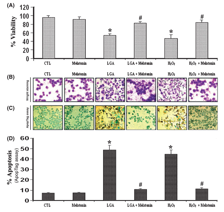
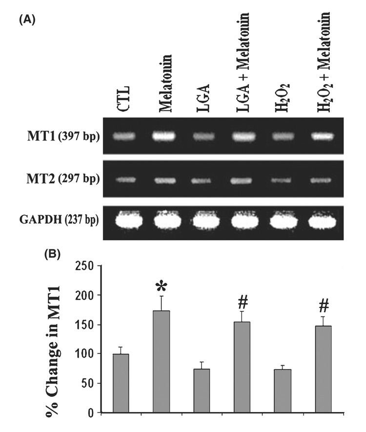
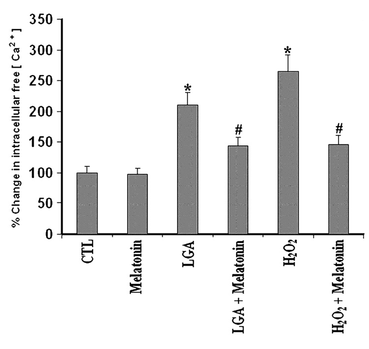
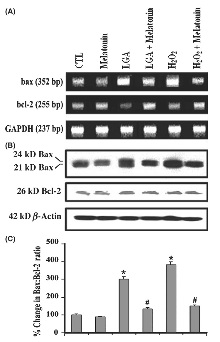
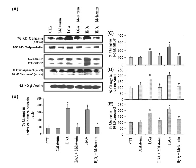
To preserve the central nervous system (CNS) function after a traumatic injury, therapeutic agents must be administered to protect neurons as well as glial cells. Cell death in CNS injuries and diseases are attributed to many factors including glutamate toxicity and oxidative stress. We examined whether melatonin, a potent anti-oxidant and free radical scavenger, would attenuate apoptotic death of rat C6 astroglial cells under glutamate excitotoxicity and oxidative stress. Exposure of C6 cells to 500 microM L-glutamic acid (LGA) and 100 microm hydrogen peroxide (H(2)O(2)) for 24 hr caused significant increases in apoptosis. Apoptosis was evaluated by Wright staining and ApopTag assay. Melatonin receptor 1 appeared to be involved in the protection of these cells from excitotoxic and oxidative damage. Cells undergoing excitotoxic and oxidative stress for 15 min were then treated with 150 nM melatonin, which prevented Ca(2+)influx and cell death. Western blot analyses showed alterations in Bax and Bcl-2 expression resulting in increased Bax:Bcl-2 ratio during apoptosis. Western blot analyses also showed increases in calpain and caspase-3 activities, which cleaved 270 kD alpha-spectrin at specific sites to generate 145 kD spectrin breakdown product (SBDP) and 120 kD SBDP, respectively. However, 15-min post-treatment of C6 cells with melatonin dramatically reduced Bax:Bcl-2 ratio and proteolytic activities, decreasing LGA or H(2)O(2)-induced apoptosis. Our data showed that melatonin prevented proteolysis and apoptosis in C6 astroglial cells. The results suggest that melatonin may be an effective cytoprotective agent against glutamate excitotoxicity and oxidative stress in CNS injuries and diseases.


Figures

Fig. 1
Fig. 1. Determination of apoptosis in C6 cells after the treatments
(A) Melatonin post-treatment prevented l-glutamic acid (LGA) or H2O2-mediated decrease in C6 cell viability. The trypan blue dye exclusion test was used to assess cell viability. (B) Wright staining showing representative cells from each treatment group. The arrows indicate apoptotic cells. (C) ApopTag assay showing representative cells from each treatment group. The arrows indicate apoptotic cells. (D) Bar graphs indicating the percentage of apoptotic cells (based on ApopTag assay). Treatment groups (panels A–D): control (CTL); 150 nm melatonin (24 hr); 500 µm LGA (24 hr); 500 µm LGA (15 min) + melatonin post-treatment (24 hr); 100 µm H2O2 (24 hr); and 100 µm H2O2 (15 min) + melatonin post-treatment (24 hr).
Fig. 2
Fig. 2. Reverse transcription-polymerase chain reaction (RT-PCR) experiments for examining levels of expression of melatonin receptors in C6 cells
(A) Representative agarose gel pictures to show levels of mRNA expression of melatonin receptor 1 (MT1) and melatonin receptor 2 (MT2). (B) Bar graphs indicating the percent change in mRNA expression of MT1. Treatment groups (panels A and B): control (CTL); 150 nm melatonin (24 hr); 500 µm LGA (24 hr); 500 µm l-glutamic acid (LGA) (15 min) + melatonin post-treatment (24 hr); 100 µm H2O2 (24 hr); and 100 µm H2O2 (15 min) + melatonin post-treatment (24 hr).
Fig. 3
Fig. 3. Determination of intracellular free [Ca2+] using fura-2
The data were from C6 cells grown in phenol-red free medium, treated for 24 hr, and then exposed to fura-2. Treatment groups: control (CTL); 150 nm melatonin (24 hr); 500 µm l-glutamic acid (LGA) (24 hr); 500 µm LGA (15 min) + melatonin post-treatment (24 hr); 100 µm H2O2 (24 hr); and 100 µm H2O2 (15 min) + melatonin post-treatment (24 hr).
Fig. 4
Fig. 4. Alterations in expression of Bax and Bcl-2 at mRNA and protein levels
(A) Representative agarose gels to show mRNA levels of bax, bcl-2, and GAPDH genes (reverse transcription-polymerase chain reaction). (B) Representative Western blots to show protein levels of Bax, Bcl-2, and GAPDH (Western blotting). (C) Densitometric analysis showing the Bax:Bcl-2 ratio in six treatment groups. Treatment groups (panels A–C): control (CTL); 150 nm melatonin (24 hr); 500 µm l-glutamic acid (LGA) (24 hr); 500 µm LGA (15 min) + melatonin post-treatment (24 hr); 100 µm H2O2 (24 hr); and 100 µm H2O2 (15 min) + melatonin post-treatment (24 hr).
Fig. 5
Fig. 5. Determination of activation and activity of calpain as well as of caspase-3
(A) Representative Western blots to show levels of 76-kD calpain active fragment, 100 kD calpastatin (endogenous calpain inhibitor), 145 kD spectrin breakdown product (SBDP), 120 kD SBDP, 32 kD caspase-3 inactive fragment and 20 kD caspase-3 active fragment, and β-actin. Densitometric analysis to show percent change (B) in active calpain: calpastatin ratio, (C) in calpain-specific 145 kD SBDP, (D) in caspase-3-specific 120 kD SBDP, and (E) in 20 kD caspase-3. Treatment groups (panels A–E): control (CTL); 150 nm melatonin (24 hr); 500 µm l-glutamic acid (LGA) (24 hr); 500 µm LGA (15 min) + melatonin post-treatment (24 hr); 100 µm H2O2 (24 hr); and 100 µm H2O2 (15 min) + melatonin post-treatment (24 hr).

References

    1. Tan DX, Manchester LC, Terron MP, et al. One molecule, many derivatives: a never-ending interaction of melatonin with reactive oxygen and nitrogen species? J Pineal Res. 2007;42:28–42. - PubMed
    1. M Hardeland R, Backhaus C, Fadavi A. Reactions of the NO redox forms NO+, *NO and HNO (protonated NO−) with the melatonin metabolite N1-acetyl-5-methoxykynuramine. J Pineal Res. 2007;43:382–388. - PubMed
    1. Mesenge C, Margaill I, Verrecchia C, et al. Protective effect of melatonin in a model of traumatic brain injury in mice. J Pineal Res. 1998;25:41–46. - PubMed
    1. León J, Acuña-Castroviejo D, Escames G, et al. Melatonin mitigates mitochondrial malfunction. J Pineal Res. 2005;38:1–9. - PubMed
    1. Jou MJ, Peng TI, Yu PZ, et al. Melatonin protects against common deletion of mitochondrial DNA-augmented mitochondrial oxidative stress and apoptosis. J Pineal Res. 2007;43:389–403. - PubMed

Publication types

MeSH terms