. 2008 Sep;125(1):111-21.
doi: 10.1111/j.1365-2567.2008.02826.x. Epub 2008 Mar 28.

The immunomodulatory glycan LNFPIII initiates alternative activation of murine macrophages in vivo

Affiliations

The immunomodulatory glycan LNFPIII initiates alternative activation of murine macrophages in vivo

Olga Atochina et al. Immunology. 2008 Sep.

Abstract

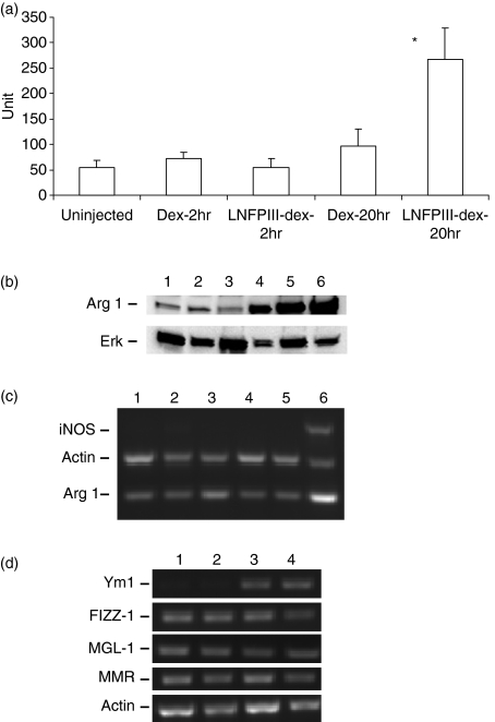
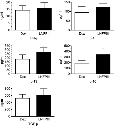
The early pathogen-macrophage interactions that help drive macrophage maturation towards classically or alternatively activated are largely unknown. To examine this question we utilized the immunomodulatory glycan Lacto-N-fucopentaose III (LNFPIII), which contains the Lewis X (LeX) trisaccharide, to activate murine peritoneal macrophages in vivo. Because LNFPIII is known to induce anti-inflammatory responses, we asked if LNFPIII stimulation of macrophages in vivo initiates alternative activation events such as upregulation of Arginase 1, Ym1, FIZZ-1, MGL-1 or macrophage mannose receptor (MMR). Examination of peritoneal exudate cells from mice 20 hr post-LNFPIII injection demonstrated increased Arginase 1 activity, at the mRNA and protein levels, coincident with undetectable inducible nitric oxide synthase expression or nitric oxide production. In addition to Arginase 1, Ym1 expression was also significantly upregulated at 20 and 48 hr after LNFPIII exposure in vivo. However, the expression of FIZZ-1, MGL-1, and MMR was not changed in these macrophages. In an attempt to determine activation requirements for functional activity, we adoptively transferred antigen-pulsed, in vivo LNFPIII activated macrophages into naïve recipients and found that they were capable of triggering recipient T cells to secrete elevated levels of interleukin (IL)-10 and IL-13 compared to mice receiving control macrophages. Together, these data demonstrate that upregulation of expression of Arginase 1 and Ym1 occur very early in activation of macrophages, and can be independent of other alternatively activated (AA) macrophage markers. Importantly, these early events appear to be IL-4/IL-13-independent in our model. In the future we hope to determine if upregulation of these initial AA maturational events is sufficient for these macrophages to exert immunoregulatory activity in vivo.


Figures

Figure 1
Figure 1
LNFPIII-dex upregulates Arginase 1 and Ym1 of PEC in vivo. Groups of five to six mice were left uninjected or injected with 50 μg of Dex or LNFPIII-dex. Mice were killed at indicated time points and peritoneal macrophages were isolated. (a) Arginase 1 activity was determined in PEC lysates from all mice as described in Materials and methods. One unit of enzyme activity is defined as the amount of enzyme that catalyzes the formation of 1 μmol urea per 1 hr. All data of four experiments are summarized and expressed as the mean ± SD. A statistical significance between groups was determined by Student's t-test (P < 0·001), and significant differences are indicated with asterisk. (b) Western blot analysis of Arginase 1 in PEC obtained from uninjected mice (lane 1), 2 hr Dex-injected mice (lane 2), 2 hr LNFPIII-dex injected mice (lane 3), 20 hr LNFPIII-dex injected mice (lane 4). PEC stimulated with 20 ng/ml rmIL-4 (lane 5) and 1 μg/ml lipopolysaccharide (LPS) + 20 ng/ml IFN-γ (lane 6) for 20 hr were used as positive controls. The same blot was stripped and reprobed with anti-ERK antibody. (c) Expression levels of Arginase 1 and iNOS were determined by multiplex RT–PCR for PEC from uninjected mice (lane 1), mice injected with Dex for 20 hr (lane 2) or 48 hr (lane 4), LNFPIII-dex for 20 (lane 3), or LNFPIII-dex for 48 hr (lane 5), positive control represents the PEC stimulated with LPS + IFN (lane 6). (d) Gene expression of Ym1, FIZZ1, MMR and MGL-1 in PEC from unjected mice (lane 1), injected with 50 μg/ml of Dex (lane 2), LNFPIII-dex for 20 hr (lane 3) or LNFPIII-dex for 48 hr (lane 4) was determined by RT–PCR. The RT–PCR figures are representative of three independent experiments.
Figure 2
Figure 2
Selective induction of maturation markers by LNFPIII-dex injection. Groups of four mice were injected with 50 μg/ml of Dex or LNFPIII-dex for 20 hr (a) and 48 hr (b). PEC were isolated and stained for F4/80, CD80, CD86, MHC II, ICAM-1, PD-L1 and ICOS-L. Expression of costimulatory molecules was analysed for gated F4/80+ cells. The thin line depicts the expression of surface markers from PEC of Dex injected mice and the thick line depicts the expression from LNFPIII-dex injected mice. At least three separate experiments with similar results were performed.
Figure 3
Figure 3
Functional activity of LNFPIII-dex stimulated macrophages in vivo. Groups of four to six mice were injected with 50 μg/ml of Dex or LNFPIII-dex for 20 hr. PEC were isolated, pooled and plated for 1 hr with 1000 nm of OVA peptide (OVA323–339). Adherent cells were collected and 1 million cells were injected i.p. to groups of four DO.11.10 mice. In 5–6 days splenocytes from individual mice were isolated and plated in the presence of OVA peptide (100 and 1000 nm) for 72 hr. Cytokines were measured by ELISA. Four independent experiments were performed. Summarized data are shown as mean ± SD. A statistical significance between groups was determined by Student's t-test (P < 0·05), and significant differences are indicated with an asterisk.
Figure 4
Figure 4
Role of IL-4 and IL-13 on LNFPIII-elicited macrophages. Groups of wild type mice (WT) or IL-4Rα KO mice were left unjected (lane 1, WT; lane 4 KO), injected with 50 μg/ml of Dex (lane 2, WT; lane 5 KO), LNFPIII-dex for 20 hr (lane 3, WT; lane 6 KO). Total RNA was isolated using TRIzol reagent, and gene expression was determined by RT–PCR for iNOS and Argiase 1 (a), and Ym1 and FIZZ-1 (b). (a) and (b) RT-PCR figures are representative of at least three independent experiments. (c) Real-time PCR was performed for Arginase 1, Ym1, and FIZZ-1 expression in PECs from wild type and IL-4Rα KO mice. Total RNA was isolated using Qiagen RNeasy Kit. GAPDH was used to normalize loading. Results of two independent experiments are summarized and presented as a fold of differences over the control (uninjected). Data are shown as mean ± standard error. (d) PEC from the same mice were stained for F4/80, CD80 and PD-L1. Expression of CD80 and PD-L1 was analysed for gated F4/80+ cells. The thin line depicts the expression of surface markers from PEC of Dex-injected mice and the thick line depicts the expression from LNFPIII-dex injected mice. The results for one representative of two independent experiments are shown.

References

    1. Nathan C, Shiloh MU. Reactive oxygen and nitrogen intermediates in the relationship between mammalian hosts and microbial pathogens. Proc Natl Acad Sci U S A. 2000;97:8841. - PMC - PubMed
    1. Stain SC, Baer HU, Dennison AR, Blumgart LH. Current management of hilar cholangiocarcinoma. Surg Gynecol Obstet. 1992;175:579. - PubMed
    1. Gordon S. Alternative activation of macrophages. Nat Rev Immunol. 2003;3:23. - PubMed
    1. Anthony RM, Urban JF, Jr, Alem F, Hamed HA, Rozo CT, Boucher JL, Van Rooijen N, Gause WC. Memory T(H)2 cells induce alternatively activated macrophages to mediate protection against nematode parasites. Nat Med. 2006;12:955. - PMC - PubMed
    1. Edwards JP, Zhang X, Frauwirth KA, Mosser DM. Biochemical and functional characterization of three activated macrophage populations. J Leukoc Biol. 2006;80:1298. - PMC - PubMed

Publication types

MeSH terms

LinkOut - more resources