. 2009 Feb;13(2):320-33.
doi: 10.1111/j.1582-4934.2008.00324.x. Epub 2008 Mar 28.

Neuronal mitochondrial amelioration by feeding acetyl-L-carnitine and lipoic acid to aged rats

Affiliations

Neuronal mitochondrial amelioration by feeding acetyl-L-carnitine and lipoic acid to aged rats

Gjumrakch Aliev et al. J Cell Mol Med. 2009 Feb.

Abstract

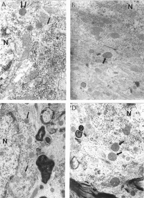
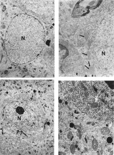
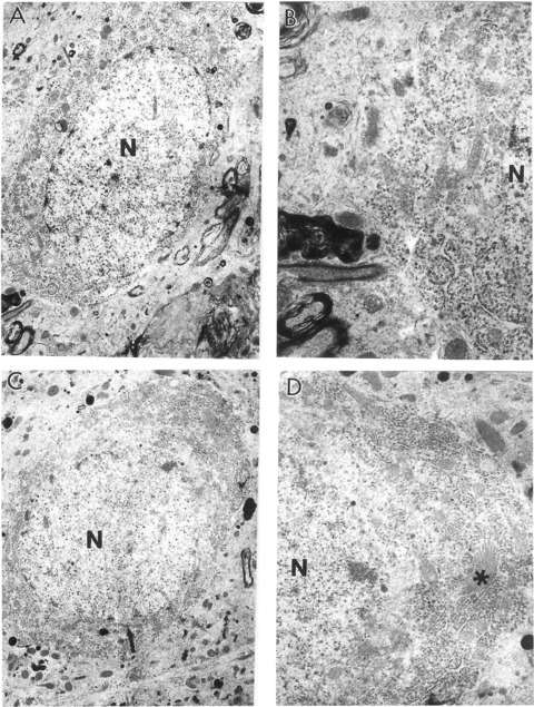
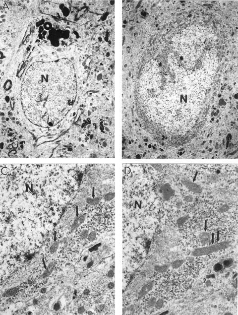
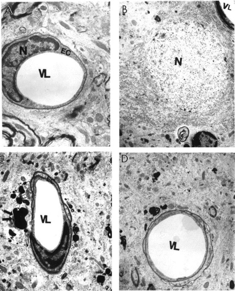
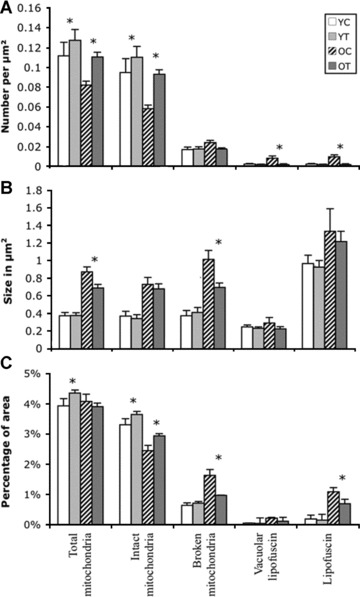
Brain function declines with age and is associated with diminishing mitochondrial integrity. The neuronal mitochondrial ultrastructural changes of young (4 months) and old (21 months) F344 rats supplemented with two mitochondrial metabolites, acetyl-L-carnitine (ALCAR, 0.2%[wt/vol] in the drinking water) and R-alpha-lipoic acid (LA, 0.1%[wt/wt] in the chow), were analysed using qualitative and quantitative electron microscopy techniques. Two independent morphologists blinded to sample identity examined and scored all electron micrographs. Mitochondria were examined in each micrograph, and each structure was scored according to the degree of injury. Controls displayed an age-associated significant decrease in the number of intact mitochondria (P = 0.026) as well as an increase in mitochondria with broken cristae (P < 0.001) in the hippocampus as demonstrated by electron microscopic observations. Neuronal mitochondrial damage was associated with damage in vessel wall cells, especially vascular endothelial cells. Dietary supplementation of young and aged animals increased the proliferation of intact mitochondria and reduced the density of mitochondria associated with vacuoles and lipofuscin. Feeding old rats ALCAR and LA significantly reduced the number of severely damaged mitochondria (P = 0.02) and increased the number of intact mitochondria (P < 0.001) in the hippocampus. These results suggest that feeding ALCAR with LA may ameliorate age-associated mitochondrial ultrastructural decay and are consistent with previous studies showing improved brain function.


Figures

1
1
Ultrastructural characteristics of hippocampal neurons in young and old rats with and without 0.2% ALCAR and 0.1% LA diet supplementation. (A) The cell body from a young control rat hippocampus shows the presence of a large number of normal mitochondria. Some of the mitochondria however show minimal changes in their matrix (single arrows indicate the presence of intramitochondrial oedema; double arrows indicate electron dense mitochondria). (B) Mitochondria from the neu-ronal cell body of YT rat show completely intact morphology (arrow indicates normal healthy mitochondrion) (×15,000). (C) The main features of neuronal abnormality in aged control rats appear to be the presence of a range of mitochondria lesions. Some mitochondria however still show intact morphology (single arrows). A giant lipofuscin deposit was close to this neuronal cell body (×15,000). (D) Hippocampal neurons from the OT group are characterized by the absence of mitochondria degeneration. Single arrows indicate normal mitochondria. Lipofuscin was also absent in the neuronal cell body as well as in other cellular compartments in this group of experiments (×15.000). N, nucleus.
2
2
Features of hippocampal neurons from old control rats. (A) A neuron under lower magnification. The neuronal cell body shows different degrees of mitochondrial abnormality (×5000). (B) A giant mitochondrion (arrow) is present in the bottom central portion of this neuron seen under high magnification. Many mitochondria are at the stage of transforming to mitochondrial-derived lysosomal structures (×15,000). (C) Another neuron under the lower magnification. A large lipofuscin deposit characterizes the cell body (X5000). (D) Neuronal cell body under high magnification. Giant mitochondria (arrows) and mitochondria transforming to mitochondrial-derived lysosomes (that appear to be part of lipofuscin formation) are consistent features in this neuron (x15,000). N, nucleus.
3
3
Features of mitochondrial abnormalities in aged OC rat hippocampi. (A) Normal mitochondria (arrows) in neuron under low magnification (×6000). (B) The cell body of this neuron under higher magnification. The cell body contains different populations of mitochondria including normal mitochondria (arrows). Microtubules coexist with normal but not damaged mitochondria (×15,000). (C) Another neuron from the hippocampal area under the low power. Double arrows indicate giant mitochondria (×5000). (D) Cell body of this neuron under higher magnification. Double asterisks indicate giant mitochondria (×15,000). N, nucleus.
4
4
The features of aged rat hippocampal astrocytes and neurons. (A) Astrocytes under low magnification (×6000). (B) High magnification image of the cell bodies of this astrocytes reveals mitochondria with electron-dense matrices (indicated by single arrow) (×10,000). (C) A neuron under low magnification. The single and double arrows indicate different sized giant mitochondria (X5000). (D) The cell body of this neuron shows the presence of oedema in the mitochondrial matrix (single arrows) (×15,000). N, nucleus.
6
6
The characteristics of hippocampal neurons from aged rats treated with 0.2% ALCAR and 0.1% LA for 3 months. (A) A neuron under low magnification (×6000). (B) The cell body of this neuron under higher magnification. Only some mitochondria show partial damage to their cristae (×15,000). (C) Other neurons from this rat. The main feature of this neuron is the absence of lipofuscin in the neuronal cell body (×5000). (D) Top portion of this neuronal cell body under higher magnification. The single asterisk indicates the Golgi apparatus. No abnormalities were seen in the Golgi structure. Additional features of this neuron appear to be well developed granular and agranular endoplasmic reticu-lum and the presence of free ribosomes in the matrix of the cell body (×10,000).N, nucleus.
5
5
The heterogeneous morphology of mitochondria in aged OC rat hippocampal neurons. (A) The presence of vacuolar structures in the matrix of nuclei appears to be a feature of non-reversibly damaged neurons. The density of the cell body is notably less than that of non-damaged neurons (×5000). (B) Neurons under low magnification. Mitochondrial damage was found adjacent to areas of lipofuscin formation (right bottom portion of this neuron) (×5000). (C and D) Cell body under the higher magnification. Aged, neuronal mitochondria are characterized by increased electron density of their matrices. Oedema appeared to be a consistent feature of these neurons (Fig. C). The cell body contains a range of mitochondrial abnormalities. Hippocampal neurons display mitochondrial density transformations (single and double arrows indicate electron dense and giant mitochondria respectively) (×15,000, C and D).
7
7
Ultrastructural characteristics of microvessels in rat hippocampus with and without ALCAR + LA dietary supplementation. (A) Capillary vessels from young control non-treated hippocampal tissues had an absence of abnormalities in their ultrastructure. Mitochondria were intact and showed no abnormalities in the vascular endothelium or perivascular cellular compartments (×8000). (B) Young treated rat hippocampal microvessels were similar to young control non-treated animals, characterized by intact morphology (×6000). (C) Old non-treated rat hippocampal capillary vessel endothelium showed degenerative changes in their ultrastructure, which appeared to be membrane disruption and nuclear contraction. A giant lipofuscin formation derived from a completely damaged mitochondrion in the matrix of a perivascular cell was also seen (double arrow) (×6000). (D) A hippocampal capillary from old ALCAR + LA treated rat. Vascular endothelial and perivascular cells showed no damage in their ultrastructure. Occasionally, a single small lipofuscin formation was present in the matrix of perivascular cells (×6000). EC, endothelial cell; N, nucleus; VL, vessel lumen.
8
8
(A) The percentage of intact mitochondria in hippocampal neurons. Abbreviations: YC, young control non-treated rats; YT, young rats treated with ALCAR + LA dietary supplementation; OC, old control rats non-treated; OT, old rats treated with ALCAR + LA. (B) The effect of ALCAR + LA dietary supplementation on the distribution of the different types of mitochondria. Significance differences are indicated: *P < 0.05, treated groups compared to respective control; #P< 0.05, significant decrease compared to young control; ##P < 0.01, increase compared to young control.
9
9
Morphometric quantification of (A) number, (B) size and (C) percentage coverage of cytoplasmic area by intact mitochondria, mitochondria with broken cristae, total mitochondria (intact plus mitochondria with broken cristae), vacuole associated lipofuscin, and lipofuscin alone in cases of young control (YC), young treated (YT), old control (OC) and old treated (OT). (A) Number of intact mitochondria is significantly lower in OC (P = 0.001). No significant change was seen in the number of mitochondria with broken cristae; however, the number of vacuoles associated with lipofuscin and lipofuscin alone were significantly higher in OC, which was significantly decreased with dietary supplementation. (B) A significant decrease in the average size of mitochondria with broken cristae was seen in old non-treated groups compared to treated animals. No significant difference was seen in the average size of intact mitochondria, lipofuscin-associated vacuoles or lipofuscin. (C) Treatment with ALCAR + LA supplementation significantly increased the percentage of area of intact mitochondria in old and young animals. However, a significant difference was seen in the percentage of area covered by mitochondria with broken cristae and lipofuscin only in OC compared to OT groups.

Similar articles

Cited by

References

    1. de Grey ADJ. The mitochondrial free radical theory of aging. Georgetown, TX: R.G. Landers Company; 1999.
    1. Hagen TM, Yowe DL, Bartholomew JC, Wehr CM, Do KL, Park JY, Ames BN. Mitochondrial decay in hepatocytes from old rats: membrane potential declines, heterogeneity and oxidants increase. Proc NatlAcad Sci USA. 1997;94:3064–9. - PMC - PubMed
    1. Shigenaga MK, Hagen TM, Ames BN. Oxidative damage and mitochondrial decay in aging. Proc Natl Acad Sci USA. 1994;91:10771–8. - PMC - PubMed
    1. Wallace DC. A mitochondrial paradigm of metabolic and degenerative diseases, aging, and cancer: a dawn for evolutionary medicine. Annu Rev Genet. 2005;39:359–407. - PMC - PubMed
    1. Sohal RS, Mockett RJ, Orr WC. Mechanisms of aging: an appraisal of the oxidative stress hypothesis. Free Radic BiolMed. 2002;33:575–86. - PubMed

Publication types