. 2008 Dec;12(6B):2675-90.
doi: 10.1111/j.1582-4934.2008.00321.x. Epub 2008 Mar 28.

Deregulation of keratinocyte differentiation and activation: a hallmark of venous ulcers

Affiliations

Deregulation of keratinocyte differentiation and activation: a hallmark of venous ulcers

Olivera Stojadinovic et al. J Cell Mol Med. 2008 Dec.

Abstract

Epidermal morphology of chronic wounds differs from that of normal epidermis. Biopsies of non-healing edges obtained from patients with venous ulcers show thick and hyperproliferative epidermis with mitosis present in suprabasal layers. This epidermis is also hyper-keratotic and parakeratotic. This suggests incomplete activation and differentiation of keratinocytes. To identify molecular changes that lead to pathogenic alterations in keratinocyte activation and differentiation pathways we isolated mRNA from non-healing edges deriving from venous ulcers patients and determined transcriptional profiles using Affymetrix chips. Obtained transcriptional profiles were compared to those from healthy, unwounded skin. As previously indicated by histology, we found deregulation of differentiation and activation markers. We also found differential regulation of signalling molecules that regulate these two processes. Early differentiation markers, keratins K1/K10 and a subset of small proline-rich proteins, along with the late differentiation marker filaggrin were suppressed, whereas late differentiation markers involucrin, transgultaminase 1 and another subset of small proline-rich proteins were induced in ulcers when compared to healthy skin. Surprisingly, desomosomal and tight junction components were also deregulated. Keratinocyte activation markers keratins K6/K16/K17 were induced. We conclude that keratinocytes at the non-healing edges of venous ulcers do not execute either activation or differentiation pathway, resulting in thick callus-like formation at the edge of a venous ulcers.


Figures
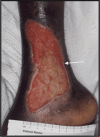
Fig. 1
Fig. 1
An image of a venous ulcer used in this study. The arrow is pointing to the location of a non-healing edge from which biopsy for microarray analysis was obtained.
Fig. 2
Fig. 2
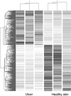
Gene tree showing different gene expression patterns between venous ulcers (left) and healthy skin (right).
Fig. 3
Fig. 3
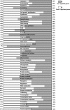
Percentage of genes differentially regulated between non-healing edges of venous ulcers and healthy skin samples sorted according to biological functions.
Fig. 4
Fig. 4
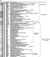
A hundred top regulated genes. Fold changes are shown in left columns. P-values, gene symbols and unigene comments are given next. The genes are marked with brackets and grouped according to cellular functions and biological processes.
Fig. 4
Fig. 4
A hundred top regulated genes. Fold changes are shown in left columns. P-values, gene symbols and unigene comments are given next. The genes are marked with brackets and grouped according to cellular functions and biological processes.
Fig. 5
Fig. 5
K17 is up-regulated in non-healing edges of venous ulcers. (A) Real-time PCR confirms K17 up-regulation in ulcers. K17 expression was normalized to expression level of HPRT. (B) Immunofluorescence with K17-specific antibody shows induction of K17 in non-healing edge of venous ulcer. Scale bar 50 μm.
Fig. 5
Fig. 5
K17 is up-regulated in non-healing edges of venous ulcers. (A) Real-time PCR confirms K17 up-regulation in ulcers. K17 expression was normalized to expression level of HPRT. (B) Immunofluorescence with K17-specific antibody shows induction of K17 in non-healing edge of venous ulcer. Scale bar 50 μm.
Fig. 6
Fig. 6
Real-time PCR confirmation of selected genes differentially expressed in healthy skin and venous ulcers. Quantitative real-time PCR for the expression of BMP2 and BMP7 (A), APOD and DEFB4 (B), CCL27 and MMP11 (C), S100A7 and KLK6 (D). Mean values of expression levels were represented after normalization to expression level of HPRT1.
Fig. 6
Fig. 6
Real-time PCR confirmation of selected genes differentially expressed in healthy skin and venous ulcers. Quantitative real-time PCR for the expression of BMP2 and BMP7 (A), APOD and DEFB4 (B), CCL27 and MMP11 (C), S100A7 and KLK6 (D). Mean values of expression levels were represented after normalization to expression level of HPRT1.
Fig. 7
Fig. 7
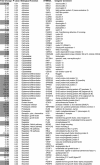
Activation and differentiation related genes. Fold changes, P-values, biological functions, gene symbols and unigene comments for genes implicated in keratinocyte activation and differentiation are shown.
Fig. 8
Fig. 8
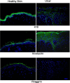
Keratinocyte differentiation markers are deregulated in venous ulcers. Immunofluorescence staining of skin specimens deriving from venous ulcers with K10, involucrin and filaggrin-specific antibodies show increased involucrin expression while K10 and filaggrin are barely detected. Scale bar 50 μm.
Fig. 9
Fig. 9
Desmosomal components are missexpressed and deregulated in venous ulcers. Immunofluorescence staining with DSC2- and DSG3-specific antibodies showed increased signal throughout the epidermis of venous ulcers while staining signal of DP was decreased. Scale bar 50 μm.

References

    1. Valencia IC, Falabella A, Kirsner RS, Eaglstein WH. Chronic venous insufficiency and venous leg ulceration. J Am Acad Dermatol. 2001;44:401–21. - PubMed
    1. Abbade LP, Lastoria S. Venous ulcer: epidemiology, physiopathology, diagnosis and treatment. Int J Dermatol. 2005;44:449–56. - PubMed
    1. Brem H, Kirsner RS, Falanga V. Protocol for the successful treatment of venous ulcers. Am J Surg. 2004;188:1–8. - PubMed
    1. Etufugh CN, Phillips TJ. Venous ulcers. Clin Dermatol. 2007;25:121–30. - PubMed
    1. Olin JW, Beusterien KM, Childs MB, Seavey C, McHugh L, Griffiths RI. Medical costs of treating venous stasis ulcers: evidence from a retrospective cohort study. Vasc Med. 1999;4:1–7. - PubMed

Publication types