. 2008 Dec;12(6B):2651-64.
doi: 10.1111/j.1582-4934.2008.00319.x. Epub 2008 Mar 28.

Sildenafil-mediated neovascularization and protection against myocardial ischaemia reperfusion injury in rats: role of VEGF/angiopoietin-1

Affiliations

Sildenafil-mediated neovascularization and protection against myocardial ischaemia reperfusion injury in rats: role of VEGF/angiopoietin-1

Srikanth Koneru et al. J Cell Mol Med. 2008 Dec.

Abstract

Sildenafil citrate (SC), a drug for erectile dysfunction, is now emerging as a cardiopulmonary drug. Our study aimed to determine a novel role of sildenafil on cardioprotection through stimulating angiogenesis during ischaemia (I) reperfusion (R) at both capillary and arteriolar levels and to examine the role of vascular endothelial growth factor (VEGF) and angiopoietin-1 (Ang-1) in this mechanistic effect. Rats were divided into: control sham (CS), sildenafil sham (SS), control+IR (CIR) and sildenafil+IR (SIR). Rats were given 0.7 mg/kg, (i.v) of SC or saline 30 min. before occlusion of left anterior descending artery followed by reperfusion (R). Sildenafil treatment increased capillary and arteriolar density followed by increased blood flow (2-fold) compared to control. Treatment with sildenafil demonstrated increased VEGF and Ang-1 mRNA after early reperfusion. PCR data were validated by Western blot analysis. Significant reduction in infarct size, cardiomyocyte and endothelial apoptosis were observed in SC-treated rats. Increased phosphorylation of Akt, eNOS and expression of anti-apoptotic protein Bcl-2, and thioredoxin, hemeoxygenase-1 were observed in SC-treated rats. Echocardiography demonstrated increased fractional shortening and ejection fraction following 45 days of reperfusion in the treatment group. Stress testing with dobutamine infusion and echocardiogram revealed increased contractile reserve in the treatment group. Our study demonstrated for the first time a strong additional therapeutic potential of sildenafil by up-regulating VEGF and Ang-1 system, probably by stimulating a cascade of events leading to neovascularization and conferring myocardial protection in in vivo I/R rat model.


Figures

Fig. 1
Fig. 1
Sildenafil treatment induced tube-like structure. Human umbilical vein endothelial cells (HUVECs) were exposed to various concentration of sildenafil ranging from 100 nM (Fig. 1B), 10 μM (Fig. 1C), 20 μM (Fig. 1D). The tube formation was very prominent when cells were exposed to 10–20 μM. VEGF siRNA (Fig. 1E) and Angiopoietin-1 siRNA (Fig. 1F) treated HUVECs abolished the tube formation induced by sildenafil.
Fig. 2
Fig. 2
Effects of sildenafil on infarct size, dp/dtmax and blood flow. (A) Bar graph shows infarct size measured 24 hrs after reperfusion. (B) Measurement of dp/dtmax under dobutamine stress test. Changes in dp/dtmax in mm of mercury/s (mmHg/s) from baseline (0 μg/kg/min) to a higher bolus dose of dobutamine (5 μg/kg/min) in 14 days I/R hearts; (C) Regional blood flow of risk area was measured with gold label neutron activation technique. Blood flow was estimated 14 days after reperfusion. Values are mean ± S.E.M. (N= 6). *P < 0.05 compared with control. CIR represents control IR and SIR represents sildenafil IR group.
Fig. 3
Fig. 3
Effect of sildenafil on apoptosis, capillary density and arteriolar density. (A) Cardiomyocyte apoptosis following 24 and 48 hrs of reperfusion. (B) shows representative pictures for cardiomyocyte apoptosis in green fluorescent colour denoted by white arrows (C) Endothelial apoptosis following 24 and 48 hrs of reperfusion. (D) Shows representative pictures for endothelial apoptosis in green fluorescent colour denoted by white arrows. (E) Left ventricular endocardial capillary density following 2 and 4 days of reperfusion. Endothelial cells were labelled using mouse monoclonal anti-CD31/PECAM-1 for capillary staining and eight non-overlapping random fields were selected from endocardial regions on non-infarcted area of the left ventricle (two sections from each heart); (F) Shows the representative pictures for capillaries stained in brown by DAB and denoted by white arrows following 4 days of reperfusion; (G) Left ventricular endocardial arteriolar density following 7 days of reperfusion. Tissue sections were labelled using monoclonal anti-smooth muscle actin and eight non-overlapping random fields were selected from endocardial region of the left ventricle; (H) Representative pictures for arterioles stained in green fluorescence and denoted by white arrows following 7 days of reperfusion. Values are mean ± S.E.M. (N= 6). *P < 0.05 compared with CIR. CIR represents control IR and SIR represents sildenafil IR group.
Fig. 4
Fig. 4
Effect of sildenafil on VEGF, Ang-1 and And-2 mRNA levels. Bar graph represents quantitative measurement of Real Time RT-PCR analysis between control I/R (CIR) and Sildenafil I/R groups following 8, 12 & 24 hrs of reperfusion: (A) VEGF mRNA; (B) Angiopoietin-1 mRNA; (C) Angiopoietin-2 mRNA *P < 0.05 compared with CIR.
Fig. 5
Fig. 5
Effect of sildenafil on phosphorylation of eNOS and Akt. A and C show a representative Western blot for the effects of sildenafil on the expression of phosphorylated-eNOS and phosphorylated-Akt in CIR and SIR following 8 hrs of reperfusion. p-eNOS and p-Akt were expressed as 140 and 60 kD respectively. eNOS and Akt were used as loading controls. Bar graphs in B and D represent the quantitative measurement in arbitrary units for p-eNOS and p-Akt respectively. *P < 0.05 compared with CIR. CIR represents control IR group and SIR represents sildenafil IR group.
Fig. 6
Fig. 6
Effect of sildenafil on the expression of HO-1, Trx-1, VEGF, Ang-1 and Bcl2. A, B, C, D and E show representative Western blots for the effects of sildenafil on the expression of (A) HO-1; (B) Trx-1; (C) VEGF; (D) Ang-1 and (E) Bcl-2 in CIR and SIR following 2, 4, 7 days of reperfusion. HO-1, Trx-1, VEGF, Ang-1 and Bcl-2 were expressed as 32, 12, 40, 60 and 26 kD respectively. GAPDH was used as the loading control. Bar graph below each Western blot shows the optical density ratio in arbitrary units. *P < 0.05 compared with CIR. CIR represents control IR group and SIR represents sildenafil IR group.
Fig. 7
Fig. 7
Echocardiographic analysis. (A) Representative pulse wave Doppler of mitral wave velocity showing improved E/A in sildenafil IR group with minimal diastolic dysfunction compared to control IR having restrictive pattern of filling. Normal diastolic function was observed in CS and SS groups. (B) M-mode of echocardiograph pictures showing improved wall motion in SIR compared to CIR and was represented by increased fractional shortening and ejection fraction in sildenafil-treated group. (C) Parasternal short axis view showing dilated chamber in CIR compared to SIR. (D) Echocardiographic measurements in control and sildenafil-treated rats following 45 days of reperfusion. (i) Ejection fraction (%), (ii) fractional shortening (%), (iii) left ventricular inner diameter (LVIDs) (mm), (iv) E/A ratio. *P < 0.05 represents significant difference between CIR and SIR groups. Significant difference was not observed in CS and SS groups. LVIDs represents left ventricular internal diameter in systole. n= 6 in each group. CS, control sham; SS, sildenafil sham; CIR, control IR group and SIR, sildenafil IR group.
Fig. 8
Fig. 8
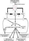
Schematic diagram. The mechanism of sildenafil-induced neovascularization in the infarcted myocardium.

References

    1. Das S, Maulik N, Das DK, Kadowitz PJ, Bivalacqua TJ. Cardioprotection with sildenafil, a selective inhibitor of cyclic 3′,5′ -monophosphate-specific phosphodi-esterase 5. Drugs Exp Clin Res. 2002;28:213–9. - PubMed
    1. Kukreja RC, Salloum F, Das A, Ockaili R, Yin C, Bremer YA, Fisher PW, Wittkamp M, Hawkins J, Chou E, Kukreja AK, Wang X, Marwaha VR, Xi L. Pharmacological preconditioning with sildenafil: basic mechanisms and clinical implications. Vascul Pharmacol. 2005;42:219–32. - PubMed
    1. Ockaili R, Salloum F, Hawkins J, Kukreja RC. Sildenafil (Viagra) induces powerful cardioprotective effect via opening of mitochondrial K(ATP) channels in rabbits. Am J Physiol Heart Circ Physiol. 2002;283:H1263–9. - PubMed
    1. Salloum F, Yin C, Xi L, Kukreja RC. Sildenafil induces delayed preconditioning through inducible nitric oxide synthase-dependent pathway in mouse heart. Circ Res. 2003;92:595–7. - PubMed
    1. Ghofrani HA, Osterloh IH, Grimminger F. Sildenafil: from angina to erectile dysfunction to pulmonary hypertension and beyond. Nat Rev Drug Discov. 2006;5:689–702. - PMC - PubMed

Publication types

MeSH terms