

The estrogen and c-Myc target gene HSPC111 is over-expressed in breast cancer and associated with poor patient outcome
- PMID: 18373870
- PMCID: PMC2397527
- DOI: 10.1186/bcr1985
The estrogen and c-Myc target gene HSPC111 is over-expressed in breast cancer and associated with poor patient outcome
Abstract
Introduction: Estrogens play a pivotal role in the initiation and progression of breast cancer. The genes that mediate these processes are not fully defined, but potentially include the known mammary oncogene MYC. Characterization of estrogen-target genes may help to elucidate further the mechanisms of estrogen-induced mitogenesis and endocrine resistance.
Methods: We used a transcript profiling approach to identify targets of estrogen and c-Myc in breast cancer cells. One previously uncharacterized gene, namely HBV pre-S2 trans-regulated protein 3 (HSPC111), was acutely upregulated after estrogen treatment or inducible expression of c-Myc, and was selected for further functional analysis using over-expression and knock-down strategies. HSPC111 expression was also analyzed in relation to MYC expression and outcome in primary breast carcinomas and published gene expression datasets.
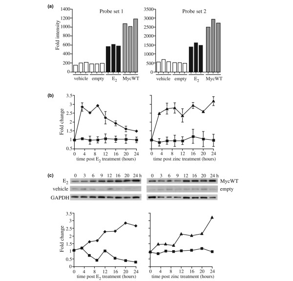
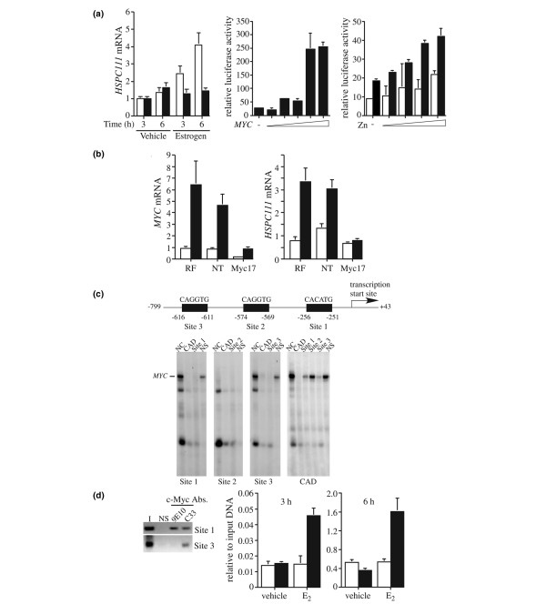
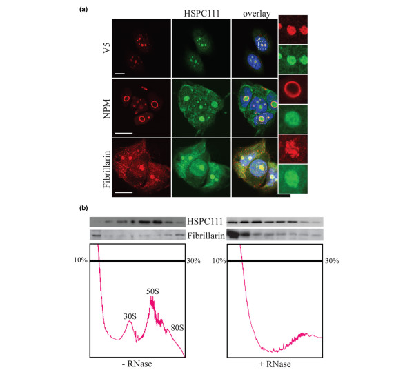
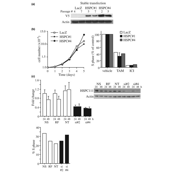
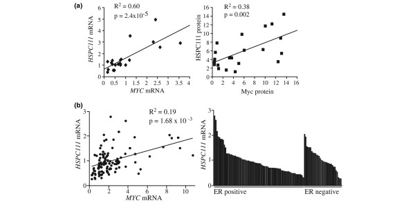
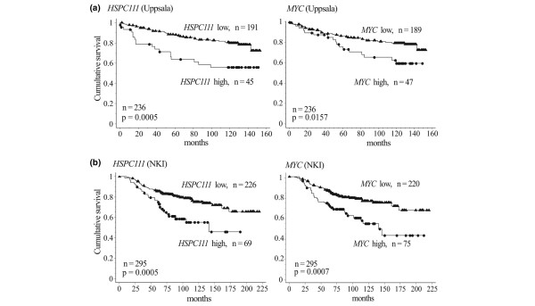
Results: Pretreatment of cells with c-Myc small interfering RNA abrogated estrogen induction of HSPC111, identifying HSPC111 as a potential c-Myc target gene. This was confirmed by the demonstration of two functional E-box motifs upstream of the transcription start site. HSPC111 mRNA and protein were over-expressed in breast cancer cell lines and primary breast carcinomas, and this was positively correlated with MYC mRNA levels. HSPC111 is present in a large, RNA-dependent nucleolar complex, suggesting a possible role in ribosomal biosynthesis. Neither over-expression or small interfering RNA knock-down of HSPC111 affected cell proliferation rates or sensitivity to estrogen/antiestrogen treatment. However, high expression of HSPC111 mRNA was associated with adverse patient outcome in published gene expression datasets.
Conclusion: These data identify HSPC111 as an estrogen and c-Myc target gene that is over-expressed in breast cancer and is associated with an adverse patient outcome.
Figures
Similar articles
-
Identification of functional networks of estrogen- and c-Myc-responsive genes and their relationship to response to tamoxifen therapy in breast cancer.PLoS One. 2008 Aug 20;3(8):e2987. doi: 10.1371/journal.pone.0002987. PLoS One. 2008. PMID: 18714337 Free PMC article.
-
c-Myc overexpression and endocrine resistance in breast cancer.J Steroid Biochem Mol Biol. 2006 Dec;102(1-5):147-55. doi: 10.1016/j.jsbmb.2006.09.028. Epub 2006 Oct 18. J Steroid Biochem Mol Biol. 2006. PMID: 17052904
-
c-Myc suppresses p21WAF1/CIP1 expression during estrogen signaling and antiestrogen resistance in human breast cancer cells.J Biol Chem. 2005 May 6;280(18):17617-25. doi: 10.1074/jbc.M502278200. Epub 2005 Mar 8. J Biol Chem. 2005. PMID: 15757889
-
Estrogen and antiestrogen regulation of cell cycle progression in breast cancer cells.Endocr Relat Cancer. 2003 Jun;10(2):179-86. doi: 10.1677/erc.0.0100179. Endocr Relat Cancer. 2003. PMID: 12790780 Review.
-
MYC in breast tumor progression.Expert Rev Anticancer Ther. 2008 Oct;8(10):1689-98. doi: 10.1586/14737140.8.10.1689. Expert Rev Anticancer Ther. 2008. PMID: 18925859 Free PMC article. Review.
Cited by
-
A pan-cancer analysis of Dyskeratosis congenita 1 (DKC1) as a prognostic biomarker.Hereditas. 2023 Dec 11;160(1):38. doi: 10.1186/s41065-023-00302-y. Hereditas. 2023. PMID: 38082360 Free PMC article.
-
Molecular characterizations of Nop16 in murine mammary tumors with varying levels of c-Myc.Transgenic Res. 2012 Apr;21(2):393-406. doi: 10.1007/s11248-011-9529-3. Epub 2011 Aug 24. Transgenic Res. 2012. PMID: 21863248 Free PMC article.
-
NOP16 promotes hepatocellular carcinoma progression and triggers EMT through the Keap1-Nrf2 signaling pathway.Technol Health Care. 2024;32(4):2463-2483. doi: 10.3233/THC-231256. Technol Health Care. 2024. PMID: 38251077 Free PMC article.
-
Identification of functional networks of estrogen- and c-Myc-responsive genes and their relationship to response to tamoxifen therapy in breast cancer.PLoS One. 2008 Aug 20;3(8):e2987. doi: 10.1371/journal.pone.0002987. PLoS One. 2008. PMID: 18714337 Free PMC article.
-
Glucose Metabolism and Glucose Transporters in Breast Cancer.Front Cell Dev Biol. 2021 Sep 6;9:728759. doi: 10.3389/fcell.2021.728759. eCollection 2021. Front Cell Dev Biol. 2021. PMID: 34552932 Free PMC article. Review.
References
-
- Colditz GA. Relationship between estrogen levels, use of hormone replacement therapy, and breast cancer. J Natl Cancer Inst. 1998;90:814–823. - PubMed
-
- Yager JD, Davidson NE. Estrogen carcinogenesis in breast cancer. N Engl J Med. 2006;354:270–282. - PubMed
-
- Sutherland RL, Hall RE, Taylor IW. Cell proliferation kinetics of MCF-7 human mammary carcinoma cells in culture and effects of tamoxifen on exponentially growing and plateau-phase cells. Cancer Res. 1983;43:3998–4006. - PubMed
-
- Taylor IW, Hodson PJ, Green MD, Sutherland RL. Effects of tamoxifen on cell cycle progression of synchronous MCF-7 human mammary carcinoma cells. Cancer Res. 1983;43:4007–4010. - PubMed
-
- Osborne CK, Boldt DH, Clark GM, Trent JM. Effects of tamoxifen on human breast cancer cell cycle kinetics: accumulation of cells in early G1 phase. Cancer Res. 1983;43:3583–3585. - PubMed
Publication types
MeSH terms
Substances
LinkOut - more resources
Full Text Sources
Medical
Molecular Biology Databases
