

Review
doi: 10.1186/bcr1871.
Epub 2008 Mar 17.
Tissue factor, angiogenesis and tumour progression
Affiliations
- PMID: 18373885
- PMCID: PMC2397518
- DOI: 10.1186/bcr1871
Item in Clipboard
Review
Tissue factor, angiogenesis and tumour progression
Breast Cancer Res.
2008.
Abstract
Tissue factor, the primary initiator of the coagulation cascade, maintains vascular integrity in response to injury. It is now recognised that, in addition to the role as a procoagulant activator, tissue factor participates in many tumour-related processes that contribute to malignant disease progression. The present review details the recent evidence supporting a role for tissue factor in tumour haemostasis, angiogenesis, metastasis and malignant cell survival. Furthermore, future research directions are discussed that may enhance our understanding of the role and regulation of this protein, which could ultimately lead to the innovative design and development of new anticancer therapies.
Figures

Tissue factor is the primary initiator of the coagulation cascade. In normal physiological conditions, following vessel damage or trauma, tissue factor (TF) forms a complex with activated factor VII (FVIIa) in the presence of calcium ions on an appropriate phospholipid membrane, and allosterically enhances the enzyme activity of this protease to catalyse the activation of factor X (FX) to FXa. Generation of FXa by the TF–FVIIa complex triggers the proteolytic conversion of prothrombin (factor II) to thrombin (activated factor II [FIIa]). This key step in the coagulation cascade is effected by the catalytic activity of the prothrombinase complex, which consists of FXa and the nonenzymatic cofactor activated factor V (FVa) in a 1:1 stoichiometric complex. The resulting burst in thrombin activity activates localised platelets and catalyses the conversion of circulating fibrinogen into an insoluble fibrin clot, thereby restoring vessel integrity.
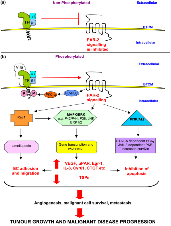
Tissue factor-induced protease-activated receptor 2 signalling. (a) When the cytoplasmic domain is nonphosphorylated, tissue factor (TF) exerts a negative regulatory control on protease-activated receptor (PAR)-2 and inhibits signalling. (b) Binding of activated factor VII (FVIIa) to TF results in proteolytic cleavage of PAR-2 and phosphorylation of the cytoplasmic domain of TF via activation of phosphatidylcholine-specific phospholipase C (PC-PLC) and protein kinase C (PKC) alpha. Phosphorylation releases the negative regulatory control of PAR-2-mediated signalling, resulting in activation of several mitogen-activated protein kinase (MAPK) pathways and subsequent gene transcription. Phosphatidylinositol 3-kinase (PI3K) and Rac1 activation also constitute important signal transduction cascades, mainly related to antiapoptotic and migration processes. The constitutive association of α3β1 integrin with aggressive breast cancer cells (for example, MDA-MB-231 cells) implies that tumour cells have lost the ability to sense extracellular gradients of coagulation proteases and constitutively couple TF and integrin signalling. Although the precise mechanism for this coupling is unknown, it may play a role in tumour cell invasiveness. The overall activation of these signalling pathways is responsible for the induction of angiogenesis, malignant cell survival and metastasis, resulting in tumour growth and disease progression. BTCM, breast tumour cell membrane; Cyr61, cysteine-rich angiogenic inducer 61; CTGF, connective tissue growth factor; EC, endothelial cell; Egr-1, early growth response gene 1; ERK, extracellular signal-related kinase; JNK, c-jun N-terminal kinase; PKB, protein kinase B; TSP, thrombospondin; uPAR, urokinase-type plasminogen activator receptor; VEGF, vascular endothelial growth factor.
References
-
- Mackman N. Alternatively spliced tissue factor – one cut too many? Thromb Haemost. 2007;97:5–8. - PubMed
Publication types
MeSH terms
Substances
LinkOut - more resources
Full Text Sources
Other Literature Sources
